Ciri dan struktur
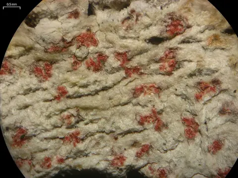
The lichen body (thallus) is a thin, whitish, crust-like growth that spreads across the bark and can disrupt the bark surface; in cross-section it is about 30–50 μm thick. The surface shows fine cracks and is bordered by brownish boundary lines (). The algal partner () is indistinct but is thought to be the green alga Trentepohlia; its cells are about 10–14 × 7 μm. The fruiting bodies (ascomata) are scattered and broadly protruding from the bark (), usually roundish to spot-like, about 0.25–1.25 mm across and 75–120 μm thick in cross-section. They sit level with the thallus and are often partly covered by bark fragments. The surface of the fruiting bodies is crimson-red and frosted with pigment (), with areas above mature spore-bearing structures (asci) turning brownish. The red pigment occurs as fine crystalline grains that can clump together into larger aggregates. Under the microscope, the uppermost tissue layer () is densely pigmented and water-repellent (hydrophobic, about 25–40 μm thick), and the asci are more or less dispersed, spherical to nearly spherical ( to subglobose, about 77–90 × 45–60 μm) with a distinct stalk. The ascospores are persistently colorless (hyaline) and divided by both transverse and longitudinal cross-walls (), measuring about 30–40 × 14–19 μm, with roughly nine transverse cross-walls and 1–3 longitudinal cross-walls per segment; the pattern of division can be irregular. In chemical reactions reported from microscopic preparations, the red pigments dissolve in KOH and shift to a purplish-orange tone, and iodine reactions are described for gels and ascus/spore structures.
Habitat dan peran ekologis
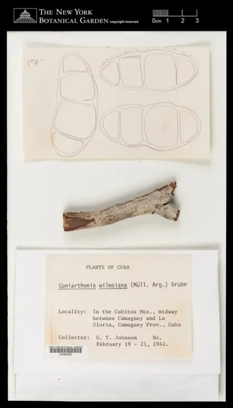
Coniarthonia gregarina is a bark-dwelling, lichen-forming species recorded from smooth bark. It was originally known from Florida based on the type and additional examined material. The lichen was later recorded from Mato Grosso do Sul, Brazil. Grube noted that it can resemble C. pyrrhula and C. wilmsiana at a glance, but it differs from both by its muriform ascospores and by having a smaller amount of reddish pigment in the ascomata; young spores with only three septa may resemble the mature spores of C. wilmsiana. Because the available material was limited, secondary chemistry was not analyzed by thin-layer chromatography.
- Habitat atau konteks temuan
- Informasi belum tersedia pada sumber rilis ini.
- Substrat
- Informasi belum tersedia pada sumber rilis ini.
- Peran atau konteks
- Pembentuk liken
Reproduksi
The fruiting bodies (ascomata) are scattered and broadly protruding from the bark (), usually roundish to spot-like, about 0.25–1.25 mm across and 75–120 μm thick in cross-section. They sit level with the thallus and are often partly covered by bark fragments. The surface of the fruiting bodies is crimson-red and frosted with pigment (), with areas above mature spore-bearing structures (asci) turning brownish. The red pigment occurs as fine crystalline grains that can clump together into larger aggregates. Under the microscope, the uppermost tissue layer () is densely pigmented and water-repellent (hydrophobic, about 25–40 μm thick), and the asci are more or less dispersed, spherical to nearly spherical ( to subglobose, about 77–90 × 45–60 μm) with a distinct stalk.
Catatan identitas dan cakupan
- Nama diterima mengikuti Catalogue of Life Base2026-09-11; uraian mempertahankan konsep dan nama sumber aslinya.
- Original geographic, specimen and life-cycle scope retained; source import awaits individual editorial review.
- Impor dan terjemahan otomatis belum diperiksa editorial per profil. Struktur reproduktif yang disebutkan tidak membuktikan seluruh siklus hidup diketahui.
- Pengukuran pada uraian mengikuti spesimen, kultur, dan metode sumber; angka tidak diperluas ke semua populasi.
Catatan distribusi dalam narasi mengikuti sumbernya. Dokumentasi spesimen atau foto mengikuti catatan sumber. Bukti wilayah Indonesia dan penilaian konservasi tidak disimpulkan dari gambar.
Melihat dengan konteks
Dokumentasi visual yang memenuhi pemeriksaan subjek dan hak penggunaan belum tersedia. Foto gejala, label, dan gambar yang meragukan tidak dihitung.
Sumber profil
- [1] Coniarthonia gregarina — Wikipedia (en)
- [2] Catalogue of Life Base2026-09-11 — Coniarthonia gregarina
Rujukan halaman ini
Fungi. Coniarthonia gregarina. Rilis data fungi-2026-10-12-expansion-2. https://fungi-atlas.mgadib.chatgpt.site/jamur/coniarthonia-gregarina/.
Status: impor sumber; pemeriksaan editorial per profil belum selesai. Untuk klaim biologis, gunakan rujukan pada bagian terkait dan periksa konteks sumber aslinya.