Ciri dan struktur
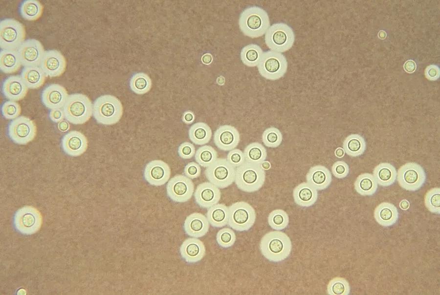
Cryptococcus neoformans typically grows as a yeast (unicellular) and replicates by budding. It makes hyphae during mating, and eventually creates basidiospores at the end of the hyphae before producing spores. Under host-relevant conditions, including low glucose, serum, 5% carbon dioxide, and low iron, among others, the cells produce a characteristic polysaccharide capsule. The recognition of C. neoformans in Gram-stained smears of purulent exudates may be hampered by the presence of the large, gelatinous capsule, which apparently prevents definitive staining of the yeast-like cells. In such stained preparations, it may appear either as round cells with Gram-positive granular inclusions impressed upon a pale lavender cytoplasmic background or as Gram-negative lipoid bodies. When grown as a yeast, C. neoformans has a prominent capsule composed mostly of polysaccharides. Under the microscope, the India ink stain is used for easy visualization of the capsule in cerebral spinal fluid. The particles of ink pigment do not enter the capsule that surrounds the spherical yeast cell, resulting in a zone of clearance or "halo" around the cells. This allows for quick and easy identification of C. neoformans. Unusual morphological forms are rarely seen. For identification in tissue, mucicarmine stain provides specific staining of polysaccharide cell wall in C. neoformans. Cryptococcal antigen from cerebrospinal fluid is thought to be the best test for diagnosis of cryptococcal meningitis in terms of sensitivity, though it might be unreliable in HIV-positive patients. The first genome sequence for a strain of C. neoformans (var. neoformans; now C. deneoformans) was published in 2005.
Habitat dan peran ekologis
Cryptococcus neoformans is an encapsulated basidiomycetous yeast belonging to the class Tremellomycetes and an obligate aerobe that can live in both plants and animals. Its teleomorph is a filamentous fungus, formerly referred to Filobasidiella neoformans. In its yeast state, it is often found in bird excrement. It has remarkable genomic plasticity and genetic variability between its strains, making treatment of the disease it causes difficult. In addition, it has been shown to cause disease in apparently immunocompetent hosts, especially in developed countries. Infection with C. neoformans is termed cryptococcosis. Most infections with C. neoformans occur in the lungs, as the fungus enters its host through the respiratory route. Because it is normally a harmless soil fungus, C. neoformans must first adapt to its new environment inside the human body, making several virulent transformations, including the formation of a polysaccharide capsule. The specific factors that enable this transformation involve sensory receptor proteins common to most soil fungi (pH sensors, carbon dioxide sensors, and intracellular iron detectors) which have been adapted to induce C. neoformans cells into rapidly becoming a dangerous, disease-causing organism.
- Habitat atau konteks temuan
- Informasi belum tersedia pada sumber rilis ini.
- Substrat
- Informasi belum tersedia pada sumber rilis ini.
- Peran atau konteks
- Belum dikategorikan pada rilis ini.
Reproduksi
Cryptococcus neoformans typically grows as a yeast (unicellular) and replicates by budding. It makes hyphae during mating, and eventually creates basidiospores at the end of the hyphae before producing spores. Under host-relevant conditions, including low glucose, serum, 5% carbon dioxide, and low iron, among others, the cells produce a characteristic polysaccharide capsule. The recognition of C. neoformans in Gram-stained smears of purulent exudates may be hampered by the presence of the large, gelatinous capsule, which apparently prevents definitive staining of the yeast-like cells. In such stained preparations, it may appear either as round cells with Gram-positive granular inclusions impressed upon a pale lavender cytoplasmic background or as Gram-negative lipoid bodies.
Catatan identitas dan cakupan
- Nama diterima mengikuti Catalogue of Life Base2026-09-11; uraian mempertahankan konsep dan nama sumber aslinya.
- Original geographic, specimen and life-cycle scope retained; source import awaits individual editorial review.
- Impor dan terjemahan otomatis belum diperiksa editorial per profil. Struktur reproduktif yang disebutkan tidak membuktikan seluruh siklus hidup diketahui.
- Pengukuran pada uraian mengikuti spesimen, kultur, dan metode sumber; angka tidak diperluas ke semua populasi.
Catatan distribusi dalam narasi mengikuti sumbernya. Dokumentasi spesimen atau foto mengikuti catatan sumber. Bukti wilayah Indonesia dan penilaian konservasi tidak disimpulkan dari gambar.
Melihat dengan konteks
Dokumentasi Cryptococcus neoformans dari Wikimedia Commons. This photomicrograph depicts Cryptococcus neoformans using a light India ink staining preparation. Life-threatening infections caused by the encapsulated fungal pathogen Cryptococcus neoformans have been increasing steadily over the past 10 years because of the onset of AIDS, and the expanded use of immunosuppressive drugs. Identifikasi mengikuti record berkas sumber.
- Jenis
- Fotografi
- Kredit
- Photo Credit: Content Providers(s): CDC/Dr. Leanor Haley
- Lisensi
- Public domain
- Sumber media
- Buka entri sumber
Sumber profil
- [1] Cryptococcus neoformans — Wikipedia (en)
- [2] Catalogue of Life Base2026-09-11 — Cryptococcus neoformans
- [3] Dokumentasi visual — Cryptococcus neoformans
Rujukan halaman ini
Fungi. Cryptococcus neoformans. Rilis data fungi-2026-10-12-expansion-2. https://fungi-atlas.mgadib.chatgpt.site/jamur/cryptococcus-neoformans/.
Status: impor sumber; pemeriksaan editorial per profil belum selesai. Untuk klaim biologis, gunakan rujukan pada bagian terkait dan periksa konteks sumber aslinya.