01 / Morfologi

Ciri dan struktur

MFLU 18-1227 Isolated from leaf spot of Lumnitzera racemosa. Culture characteristic: Colonies on MEA reaching 5–6 cm diameter after 2 days at room temperature, colonies circular to irregular, medium dense, flat or effuse, slightly raised, with edge fimbriate, fluffy to fairly fluffy, white to grey from above, light yellow to green from below; not producing pigments in agar. Asexual morph: Conidiogenous cells (8–)8.5–14 × 0.6–1.4(–1.6) μm (x ‒ = 8.4 × 1.4, n = 15), blastic, enteroblastic, flask-shaped, phialidic, hyaline and smooth-walled. Conidia (3.7–)4–4.5 × 1–1.3(–1.5) µm (x ‒ = 4 × 1.2 µm, n = 30), unicellular, subcylindrical, hyaline, smooth-walled. Based on the multigene phylogeny, Cytospora lumnitzericola is closely related to Cytospora thailandica (Fig. 1). Although conidial sizes of both species are similar, they have significant differences in nucleotides: ITS (26 nt), ACT (22 nt), and RPB2 (53 nt) (Table 5). The phylogeny derived from the ITS regions depicts C. lumnitzericola as an independent lineage close to C. brevispora CBS 116829 and C. eucalyptina CMW5882 (Fig. 2). In future, more collections are needed to confirm whether C. lumnitzericola can exist as a saprobe or endophyte as well as performing tests to confirm its pathogenicity.

Sebagian uraian ditampilkan dalam bahasa sumber; terjemahannya belum tersedia atau belum lolos pemeriksaan.

Rujukan: [1]

Pengukuran mengikuti objek, spesimen atau kultur, dan metode yang disebutkan sumber. Baca uraian asli untuk konteks lengkap.

02 / Kehidupan

Habitat dan peran ekologis

MFLU 18-1227 Isolated from leaf spot of Lumnitzera racemosa. Culture characteristic: Colonies on MEA reaching 5–6 cm diameter after 2 days at room temperature, colonies circular to irregular, medium dense, flat or effuse, slightly raised, with edge fimbriate, fluffy to fairly fluffy, white to grey from above, light yellow to green from below; not producing pigments in agar. Asexual morph: Conidiogenous cells (8–)8.5–14 × 0.6–1.4(–1.6) μm (x ‒ = 8.4 × 1.4, n = 15), blastic, enteroblastic, flask-shaped, phialidic, hyaline and smooth-walled. Conidia (3.7–)4–4.5 × 1–1.3(–1.5) µm (x ‒ = 4 × 1.2 µm, n = 30), unicellular, subcylindrical, hyaline, smooth-walled.

Sebagian uraian ditampilkan dalam bahasa sumber; terjemahannya belum tersedia atau belum lolos pemeriksaan.

Rujukan: [1]

Habitat atau konteks temuan
Informasi belum tersedia pada sumber rilis ini.
Substrat
Informasi belum tersedia pada sumber rilis ini.
Peran atau konteks
Belum dikategorikan pada rilis ini.
Temukan hubungan serupa
03 / Siklus hidup

Reproduksi

MFLU 18-1227 Isolated from leaf spot of Lumnitzera racemosa. Culture characteristic: Colonies on MEA reaching 5–6 cm diameter after 2 days at room temperature, colonies circular to irregular, medium dense, flat or effuse, slightly raised, with edge fimbriate, fluffy to fairly fluffy, white to grey from above, light yellow to green from below; not producing pigments in agar. Asexual morph: Conidiogenous cells (8–)8.5–14 × 0.6–1.4(–1.6) μm (x ‒ = 8.4 × 1.4, n = 15), blastic, enteroblastic, flask-shaped, phialidic, hyaline and smooth-walled. Conidia (3.7–)4–4.5 × 1–1.3(–1.5) µm (x ‒ = 4 × 1.2 µm, n = 30), unicellular, subcylindrical, hyaline, smooth-walled. Based on the multigene phylogeny, Cytospora lumnitzericola is closely related to Cytospora thailandica (Fig. 1).

Sebagian uraian ditampilkan dalam bahasa sumber; terjemahannya belum tersedia atau belum lolos pemeriksaan.

Rujukan: [1]

04 / Batas informasi

Catatan identitas dan cakupan

  • Nama diterima mengikuti Catalogue of Life Base2026-09-11; uraian mempertahankan konsep dan nama sumber aslinya.
  • Preserve original treatment localities, hosts and substrate scope. Materials examined describe documented specimens, not the full global range.
  • Impor dan terjemahan otomatis belum diperiksa editorial per profil. Struktur reproduktif yang disebutkan tidak membuktikan seluruh siklus hidup diketahui.
  • Pengukuran pada uraian mengikuti spesimen, kultur, dan metode sumber; angka tidak diperluas ke semua populasi.

Catatan distribusi dalam narasi mengikuti sumbernya. Dokumentasi spesimen atau foto mengikuti catatan sumber. Bukti wilayah Indonesia dan penilaian konservasi tidak disimpulkan dari gambar.

05 / Dokumentasi

Melihat dengan konteks

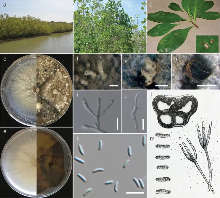
Cytospora lumnitzericola (MFLUCC 17-0508, from culture). a Mangrove collecting site b, c Lumnitzera racemosa in mangroves forest d, e Colonies on MEA after 6 days (left) and 30 days (right) (d-from above, e-from below) f, g Conidiomata produced on MEA h, l Transverse sections of conidioma i, j, n Conidiogenous cells with attached conidia k, m Conidia. Scale bars: f = 1000 µm, g, h = 500 µm, i, j = 10 µm, k = 5 µm.

Jenis
Pelat ilmiah
Kredit
Chada Norphanphoun, Olivier Raspé, Rajesh Jeewon, Ting-Chi Wen, Kevin D. Hyde
Lisensi
CC BY 4.0
Sumber media
Buka entri sumber

Gambar menunjukkan objek menurut identifikasi sumber, bukan verifikasi independen spesimen oleh Fungi. Tidak ada skala baru yang ditambahkan.

06 / Rujukan

Sumber profil

  1. [1] Morphological and phylogenetic characterisation of novel Cytospora species associated with mangroves — Cytospora lumnitzericolaBagian sumber: PMC6160782_section_8
    Tanggal penghimpunan sumber: 2026-10-12
    Teks sumber: CC BY 4.0 · Norphanphoun C, Raspé O, Jeewon R, Wen TC, Hyde KD.
  2. [2] Catalogue of Life Base2026-09-11 — Cytospora lumnitzericolaBagian sumber: ColDP NameUsage.tsv ID 6C9LT; accepted species; source 2073
    Tanggal penghimpunan sumber: 2026-10-12
  3. [3] Dokumentasi visual — Cytospora lumnitzericolaBagian sumber: Individual media record; identification follows the source, not independent determination
    Tanggal penghimpunan sumber: 2026-10-12
    Media sumber: CC BY 4.0 · Chada Norphanphoun, Olivier Raspé, Rajesh Jeewon, Ting-Chi Wen, Kevin D. Hyde

Adaptasi teks: CC BY 4.0. Norphanphoun C, Raspé O, Jeewon R, Wen TC, Hyde KD. — Morphological and phylogenetic characterisation of novel Cytospora species associated with mangroves — Cytospora lumnitzericola

Uraian asli dan riwayat sumber

Riwayat atau halaman sumber

Ringkasan sumber

MFLU 18-1227 Isolated from leaf spot of Lumnitzera racemosa. Culture characteristic: Colonies on MEA reaching 5–6 cm diameter after 2 days at room temperature, colonies circular to irregular, medium dense, flat or effuse, slightly raised, with edge fimbriate, fluffy to fairly fluffy, white to grey from above, light yellow to green from below; not producing pigments in agar. Asexual morph: Conidiogenous cells (8–)8.5–14 × 0.6–1.4(–1.6) μm (x ‒ = 8.4 × 1.4, n = 15), blastic, enteroblastic, flask-shaped, phialidic, hyaline and smooth-walled.

Revisi sumber

Morfologi sumber

MFLU 18-1227 Isolated from leaf spot of Lumnitzera racemosa. Culture characteristic: Colonies on MEA reaching 5–6 cm diameter after 2 days at room temperature, colonies circular to irregular, medium dense, flat or effuse, slightly raised, with edge fimbriate, fluffy to fairly fluffy, white to grey from above, light yellow to green from below; not producing pigments in agar. Asexual morph: Conidiogenous cells (8–)8.5–14 × 0.6–1.4(–1.6) μm (x ‒ = 8.4 × 1.4, n = 15), blastic, enteroblastic, flask-shaped, phialidic, hyaline and smooth-walled. Conidia (3.7–)4–4.5 × 1–1.3(–1.5) µm (x ‒ = 4 × 1.2 µm, n = 30), unicellular, subcylindrical, hyaline, smooth-walled. Based on the multigene phylogeny, Cytospora lumnitzericola is closely related to Cytospora thailandica (Fig. 1). Although conidial sizes of both species are similar, they have significant differences in nucleotides: ITS (26 nt), ACT (22 nt), and RPB2 (53 nt) (Table 5). The phylogeny derived from the ITS regions depicts C. lumnitzericola as an independent lineage close to C. brevispora CBS 116829 and C. eucalyptina CMW5882 (Fig. 2). In future, more collections are needed to confirm whether C. lumnitzericola can exist as a saprobe or endophyte as well as performing tests to confirm its pathogenicity.

Revisi sumber

Ekologi sumber

MFLU 18-1227 Isolated from leaf spot of Lumnitzera racemosa. Culture characteristic: Colonies on MEA reaching 5–6 cm diameter after 2 days at room temperature, colonies circular to irregular, medium dense, flat or effuse, slightly raised, with edge fimbriate, fluffy to fairly fluffy, white to grey from above, light yellow to green from below; not producing pigments in agar. Asexual morph: Conidiogenous cells (8–)8.5–14 × 0.6–1.4(–1.6) μm (x ‒ = 8.4 × 1.4, n = 15), blastic, enteroblastic, flask-shaped, phialidic, hyaline and smooth-walled. Conidia (3.7–)4–4.5 × 1–1.3(–1.5) µm (x ‒ = 4 × 1.2 µm, n = 30), unicellular, subcylindrical, hyaline, smooth-walled.

Revisi sumber

Struktur reproduksi sumber

MFLU 18-1227 Isolated from leaf spot of Lumnitzera racemosa. Culture characteristic: Colonies on MEA reaching 5–6 cm diameter after 2 days at room temperature, colonies circular to irregular, medium dense, flat or effuse, slightly raised, with edge fimbriate, fluffy to fairly fluffy, white to grey from above, light yellow to green from below; not producing pigments in agar. Asexual morph: Conidiogenous cells (8–)8.5–14 × 0.6–1.4(–1.6) μm (x ‒ = 8.4 × 1.4, n = 15), blastic, enteroblastic, flask-shaped, phialidic, hyaline and smooth-walled. Conidia (3.7–)4–4.5 × 1–1.3(–1.5) µm (x ‒ = 4 × 1.2 µm, n = 30), unicellular, subcylindrical, hyaline, smooth-walled. Based on the multigene phylogeny, Cytospora lumnitzericola is closely related to Cytospora thailandica (Fig. 1).

Revisi sumber

Rujukan halaman ini

Fungi. Cytospora lumnitzericola. Rilis data fungi-2026-10-12-expansion-2. https://fungi-atlas.mgadib.chatgpt.site/jamur/cytospora-lumnitzericola/.

Status: impor sumber; pemeriksaan editorial per profil belum selesai. Untuk klaim biologis, gunakan rujukan pada bagian terkait dan periksa konteks sumber aslinya.