Ciri dan struktur
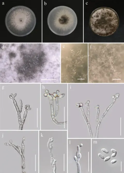
Sexual morph. Not observed. Asexual morph. Colonies effuse, hairy, dark brown to black, beige to pale brown when sporulating; vegetative hyphae immersed or semi-immersed, brown. Conidiophores (80–)118–250(–270) × (3.5–)4–5 µm, often inflated at the base up to 8–11 µm wide, macronematous, mononematous, solitary, erect, straight or slightly flexuous, cylindrical, unbranched, elongating percurrently, bearing a visible frill, brown, lower part usually dark brown and thick-walled, upper part pale brown, thinner-walled, smooth-walled, septate. Conidiogenous cells 26–47(–51) × 4.5–5.5 µm, integrated, terminal, sometimes intercalary resulting from the formation of a septum within the original cell, polyblastic, occasionally monoblastic, proliferating sympodially forming a rachis with numerous minute pimple-like denticles or protrusions scattered over the surface, cylindrical, tapering or slightly inflated in the upper part, pale brown, subhyaline towards the apex, smooth-walled; conidiogenesis holoblastic-denticulate. Conidia 10–18 × 3.5–6 µm (mean ± SD = 13.8 ± 2.0 × 4.8 ± 0.7 μm), solitary, dry, acropleurogenous, ellipsoidal, obovoid to subclavate, straight, rounded at the apex, tapering towards the base, with a minute basal scar, hyaline to subhyaline, very pale brown in mass, (0–)3(–4)-septate, mucoid sheath is inconspicuous, ephemeral, disintegrates as the conidium matures and remains collapsed on the conidial wall giving the conidia a longitudinally crumpled appearance, ornamentation absent at the apex; conidial secession schizolytic. On CMD colonies 46–48 mm diam., circular, flat, margin entire, mucoid, glossy, pale pink-orange, pale yellow pigment diffusing into the agar, reverse orange.
Habitat dan peran ekologis
Sexual morph. Not observed. Asexual morph. Colonies effuse, hairy, dark brown to black, beige to pale brown when sporulating; vegetative hyphae immersed or semi-immersed, brown. Conidiophores (80–)118–250(–270) × (3.5–)4–5 µm, often inflated at the base up to 8–11 µm wide, macronematous, mononematous, solitary, erect, straight or slightly flexuous, cylindrical, unbranched, elongating percurrently, bearing a visible frill, brown, lower part usually dark brown and thick-walled, upper part pale brown, thinner-walled, smooth-walled, septate. Conidiogenous cells 26–47(–51) × 4.5–5.5 µm, integrated, terminal, sometimes intercalary resulting from the formation of a septum within the original cell, polyblastic, occasionally monoblastic, proliferating sympodially forming a rachis with numerous minute pimple-like denticles or protrusions scattered over the surface, cylindrical, tapering or slightly inflated in the upper part, pale brown, subhyaline towards the apex, smooth-walled; conidiogenesis holoblastic-denticulate. Conidia 10–18 × 3.5–6 µm (mean ± SD = 13.8 ± 2.0 × 4.8 ± 0.7 μm), solitary, dry, acropleurogenous, ellipsoidal, obovoid to subclavate, straight, rounded at the apex, tapering towards the base, with a minute basal scar, hyaline to subhyaline, very pale brown in mass, (0–)3(–4)-septate, mucoid sheath is inconspicuous, ephemeral, disintegrates as the conidium matures and remains collapsed on the conidial wall giving the conidia a longitudinally crumpled appearance, ornamentation absent at the apex; conidial secession schizolytic.
- Habitat atau konteks temuan
- Informasi belum tersedia pada sumber rilis ini.
- Substrat
- Informasi belum tersedia pada sumber rilis ini.
- Peran atau konteks
- Belum dikategorikan pada rilis ini.
Reproduksi
Sexual morph. Not observed. Asexual morph. Colonies effuse, hairy, dark brown to black, beige to pale brown when sporulating; vegetative hyphae immersed or semi-immersed, brown. Conidiophores (80–)118–250(–270) × (3.5–)4–5 µm, often inflated at the base up to 8–11 µm wide, macronematous, mononematous, solitary, erect, straight or slightly flexuous, cylindrical, unbranched, elongating percurrently, bearing a visible frill, brown, lower part usually dark brown and thick-walled, upper part pale brown, thinner-walled, smooth-walled, septate. Conidiogenous cells 26–47(–51) × 4.5–5.5 µm, integrated, terminal, sometimes intercalary resulting from the formation of a septum within the original cell, polyblastic, occasionally monoblastic, proliferating sympodially forming a rachis with numerous minute pimple-like denticles or protrusions scattered over the surface, cylindrical, tapering or slightly inflated in the upper part, pale brown, subhyaline towards the apex, smooth-walled; conidiogenesis holoblastic-denticulate.
Catatan identitas dan cakupan
- Nama diterima mengikuti Catalogue of Life Base2026-09-11; uraian mempertahankan konsep dan nama sumber aslinya.
- Preserve original treatment localities, hosts and substrate scope. Materials examined describe documented specimens, not the full global range.
- Impor dan terjemahan otomatis belum diperiksa editorial per profil. Struktur reproduktif yang disebutkan tidak membuktikan seluruh siklus hidup diketahui.
- Pengukuran pada uraian mengikuti spesimen, kultur, dan metode sumber; angka tidak diperluas ke semua populasi.
Catatan distribusi dalam narasi mengikuti sumbernya. Dokumentasi spesimen atau foto mengikuti catatan sumber. Bukti wilayah Indonesia dan penilaian konservasi tidak disimpulkan dari gambar.
Melihat dengan konteks
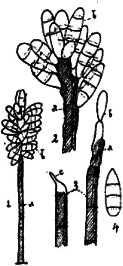
Illustration of conidiophores, conidiogenous cells and conidia of Pleurophragmium parvisporum (as P. bicolor, Costantin 1888).
- Jenis
- Pelat ilmiah
- Kredit
- Martina Réblová, Jana Nekvindová, Lucie Bauchová, Margarita Hernández-Restrepo
- Lisensi
- CC BY 4.0
- Sumber media
- Buka entri sumber
Dokumentasi tambahan
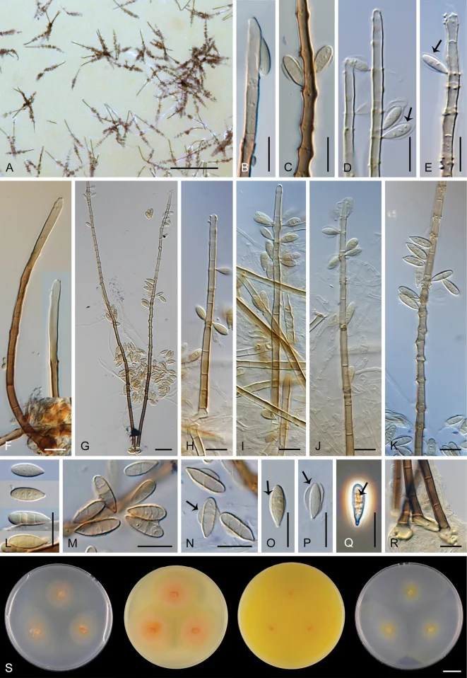
Pleurophragmium parvisporum. A Sporulating conidiophores B–E upper part of conidiophores with terminal and intercalary conidiogenous cells (nodulose swellings) and conidia (arrows indicate mucoid sheath covering the upper two-thirds of the conidium) F–K conidiophores with conidiogenous cells and conidia L–P conidia (in N, O arrows indicate mucoid wing-like sheath; in P arrow indicates mucoid sheath that disintegrates in the upper part and detaches from the wall Q conidium with longitudinally crumpled wall (arrow indicates the original collapsed sheath which causes the ornamentation) R basal cells of conidiophores S diversity of colony morphology on CMD, MLA, OA, and PCA, respectively (from left to right) after 4 wk. Images: CBS 770.83 on OA (A–E, G–K, M–Q); CBS H-3522 on natural substrate (F, L). Scale bars: 300 µm (A); 20 µm (G, R); 10 µm (B–F, H–Q); 1 cm (S).
Kredit dan penggunaan
Martina Réblová, Jana Nekvindová, Lucie Bauchová, Margarita Hernández-Restrepo
Ukuran dan format gambar disesuaikan tanpa pemotongan, retus, atau penambahan skala. Lisensi sumber dipertahankan.
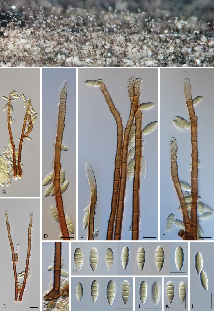
Pleurophragmium parvisporum (PRA -24032). A Sporulating conidiophores B–F conidiophores with conidiogenous cells and conidia G Basal cell H–L conidia. Images: on natural substrate. Scale bars: 300 µm (A); 20 µm (B, C); 10 µm (D–L).
Kredit dan penggunaan
Martina Réblová, Jana Nekvindová, Lucie Bauchová, Margarita Hernández-Restrepo
Ukuran dan format gambar disesuaikan tanpa pemotongan, retus, atau penambahan skala. Lisensi sumber dipertahankan.
Sumber profil
- [1] Pleurophragmium parvisporum (Ascomycota): One name, seven stories – a case highlighting the need for verification of strains from public culture collections — Pleurophragmium parvisporum
- [2] Catalogue of Life Base2026-09-11 — Dactylaria parvispora
- [3] Dokumentasi visual — Dactylaria parvispora
- [4] Dokumentasi visual — Dactylaria parvispora
- [5] Dokumentasi visual — Dactylaria parvispora
Rujukan halaman ini
Fungi. Dactylaria parvispora. Rilis data fungi-2026-10-12-expansion-2. https://fungi-atlas.mgadib.chatgpt.site/jamur/dactylaria-parvispora/.
Status: impor sumber; pemeriksaan editorial per profil belum selesai. Untuk klaim biologis, gunakan rujukan pada bagian terkait dan periksa konteks sumber aslinya.