Ciri dan struktur
Saprobic on decaying stalk of Rubus parvifolius and Hypericum monogynum. Sexual morph: Ascomata 360–440 μm high × 425–500 μm diam. (x̄ = 396 × 460 μm, n = 10), mostly gregarious, black, globose to subglobose, superficial, ostiolate. Ostiole central, minute papillate, filled with hyaline cells. Peridium 30–60 μm thick (x̄ = 47 μm, n = 30), irregularly multi-layered, comprising brown to black cells of textura angularis, with inner layer composed of flattened, hyaline cells of textura angularis. Hamathecium composed of 1–2.5 μm (x̄ = 1.7 μm, n = 30) wide, septate, hyaline, branched pseudoparaphyses. Asci (165–)180–223(–232) × (18–)19–25(–26) μm (x̄ = 200 × 22 μm, n = 20, SD = 22 × 3.3), eight-spored, bitunicate, fissitunicate, cylindrical, pedicellate, apically rounded, thick-walled at apex, with a minute ocular chamber. Ascospores (27–)29–33(–34) × (9–)10.2–12.6(–14.5) μm (x̄ = 30.8 × 11.4 μm, n = 30, SD = 2 × 1.2), muriform, with 3–7 transverse septa, and 1–3 vertical septa, with transverse septum appearing first, then vertical septa gradually emerge, mostly ellipsoidal or fusiform, rounded at both ends, initially hyaline, becoming dark brown at maturity, constricted at septa, smooth-walled, with a mucilaginous sheath. Asexual morph: Conidiomata 240–360 μm high × 185–245 µm diam (x̄ = 279 × 214 μm, n = 10), pycnidial, solitary or gregarious, superficial, obpyriform, dark brown to black, ostiolate. Ostiole 122–134 μm high × 57–62 µm wide (x̄ = 125 × 60 μm, n = 5), single, centric, circular, with hyaline periphyses, ostiolate, e single, circular, centrally papillate with or without periphyses.
Habitat dan peran ekologis
Saprobic on decaying stalk of Rubus parvifolius and Hypericum monogynum. Sexual morph: Ascomata 360–440 μm high × 425–500 μm diam. (x̄ = 396 × 460 μm, n = 10), mostly gregarious, black, globose to subglobose, superficial, ostiolate. Ostiole central, minute papillate, filled with hyaline cells. Peridium 30–60 μm thick (x̄ = 47 μm, n = 30), irregularly multi-layered, comprising brown to black cells of textura angularis, with inner layer composed of flattened, hyaline cells of textura angularis. Hamathecium composed of 1–2.5 μm (x̄ = 1.7 μm, n = 30) wide, septate, hyaline, branched pseudoparaphyses. Asci (165–)180–223(–232) × (18–)19–25(–26) μm (x̄ = 200 × 22 μm, n = 20, SD = 22 × 3.3), eight-spored, bitunicate, fissitunicate, cylindrical, pedicellate, apically rounded, thick-walled at apex, with a minute ocular chamber. Ascospores (27–)29–33(–34) × (9–)10.2–12.6(–14.5) μm (x̄ = 30.8 × 11.4 μm, n = 30, SD = 2 × 1.2), muriform, with 3–7 transverse septa, and 1–3 vertical septa, with transverse septum appearing first, then vertical septa gradually emerge, mostly ellipsoidal or fusiform, rounded at both ends, initially hyaline, becoming dark brown at maturity, constricted at septa, smooth-walled, with a mucilaginous sheath.
- Habitat atau konteks temuan
- Informasi belum tersedia pada sumber rilis ini.
- Substrat
- Informasi belum tersedia pada sumber rilis ini.
- Peran atau konteks
- Pengurai
Reproduksi
Saprobic on decaying stalk of Rubus parvifolius and Hypericum monogynum. Sexual morph: Ascomata 360–440 μm high × 425–500 μm diam. (x̄ = 396 × 460 μm, n = 10), mostly gregarious, black, globose to subglobose, superficial, ostiolate. Ostiole central, minute papillate, filled with hyaline cells. Peridium 30–60 μm thick (x̄ = 47 μm, n = 30), irregularly multi-layered, comprising brown to black cells of textura angularis, with inner layer composed of flattened, hyaline cells of textura angularis. Hamathecium composed of 1–2.5 μm (x̄ = 1.7 μm, n = 30) wide, septate, hyaline, branched pseudoparaphyses. Asci (165–)180–223(–232) × (18–)19–25(–26) μm (x̄ = 200 × 22 μm, n = 20, SD = 22 × 3.3), eight-spored, bitunicate, fissitunicate, cylindrical, pedicellate, apically rounded, thick-walled at apex, with a minute ocular chamber.
Catatan identitas dan cakupan
- Nama diterima mengikuti Catalogue of Life Base2026-09-11; uraian mempertahankan konsep dan nama sumber aslinya.
- Preserve original treatment localities, hosts and substrate scope. Materials examined describe documented specimens, not the full global range.
- Impor dan terjemahan otomatis belum diperiksa editorial per profil. Struktur reproduktif yang disebutkan tidak membuktikan seluruh siklus hidup diketahui.
- Pengukuran pada uraian mengikuti spesimen, kultur, dan metode sumber; angka tidak diperluas ke semua populasi.
Catatan distribusi dalam narasi mengikuti sumbernya. Dokumentasi spesimen atau foto mengikuti catatan sumber. Bukti wilayah Indonesia dan penilaian konservasi tidak disimpulkan dari gambar.
Melihat dengan konteks
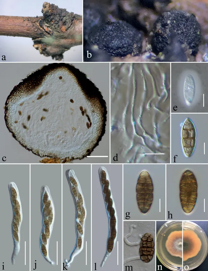
Sexual morph of Dematiomelanomma yunnanense (HKAS 124667) on decaying stalk of Rubus parvifolius L. a, b ascomata in face view c vertical section of the ascoma d pseudoparaphyses e an ascospore in Indian Ink to show a sheath f–h ascospores i–l Asci m germinating ascospore n, o surface and reverse of colony on PDA. Scale bars: 100 μm (c); 10 μm (d–h); 50 μm (i–l).
- Jenis
- Pelat ilmiah
- Kredit
- Ying Gao, Tingfang Zhong, Jayarama D. Bhat, Antonio Roberto Gomes de Farias, Turki M. Dawoud, Kevin D. Hyde, Weiqiang Xiong, Yunju Li, Heng Gui, Xuefei Yang, Shixi Wu, Dhanushka N. Wanasinghe
- Lisensi
- CC BY 4.0
- Sumber media
- Buka entri sumber
Dokumentasi tambahan
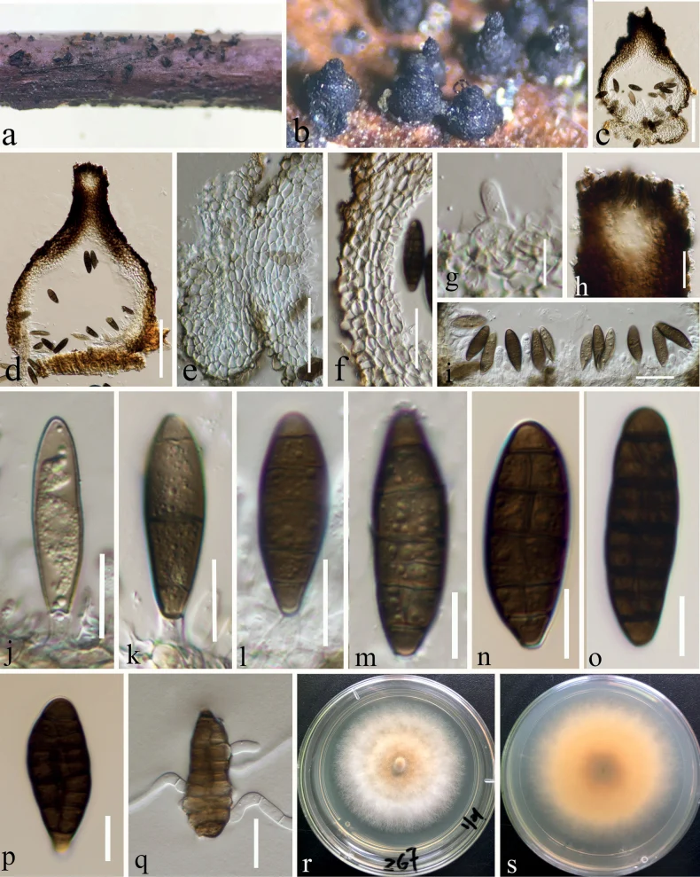
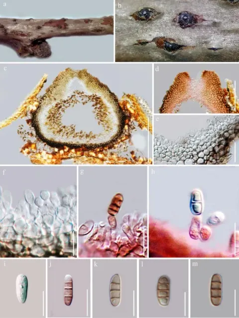
Asexual morph of Dematiomelanomma yunnanense on a dead stalk of Hypericum monogynum L. (HKAS 124666, holotype) a, b conidiomata in face view c, d vertical section of conidiomata e vertical section of the base of the pulvinate-structure f conidioma wall g conidiogenous cells arising from the wall and developing conidia h vertical section through ostiole i developing stages of conidia j–p conidia q geminating conidia r cultures on PDA from above s cultures on PDA from reverse. Scale bars: 100 μm (c, d); 50 μm (e); 30 μm (f); 10 μm (g); 20 μm (h); 30 μm (i); 15 μm (j–l); 10 μm (m–p); 20 μm (q).
Kredit dan penggunaan
Ying Gao, Tingfang Zhong, Jayarama D. Bhat, Antonio Roberto Gomes de Farias, Turki M. Dawoud, Kevin D. Hyde, Weiqiang Xiong, Yunju Li, Heng Gui, Xuefei Yang, Shixi Wu, Dhanushka N. Wanasinghe
Ukuran dan format gambar disesuaikan tanpa pemotongan, retus, atau penambahan skala. Lisensi sumber dipertahankan.
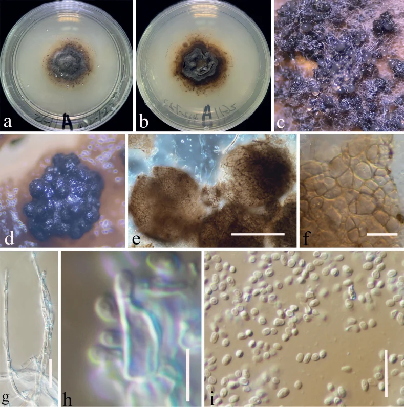
Asexual morph of Dematiomelanomma yunnanense from the culture (CGMCC 3.23744) on PDA a, b colony of the sexual morphic stage after 24 weeks on PDA (b from the bottom) c–e conidiomata f conidioma wall g mycelium h conidiogenous cells arising from the wall and developing conidia i conidia. Scale bars: 100 μm (e); 15 μm (f); 30 μm (g); 5 μm (h); 15 μm (i).
Kredit dan penggunaan
Ying Gao, Tingfang Zhong, Jayarama D. Bhat, Antonio Roberto Gomes de Farias, Turki M. Dawoud, Kevin D. Hyde, Weiqiang Xiong, Yunju Li, Heng Gui, Xuefei Yang, Shixi Wu, Dhanushka N. Wanasinghe
Ukuran dan format gambar disesuaikan tanpa pemotongan, retus, atau penambahan skala. Lisensi sumber dipertahankan.
Sumber profil
- [1] Pleomorphic Dematiomelanomma yunnanense gen. et sp. nov. (Ascomycota, Melanommataceae) from grassland vegetation in Yunnan, China — Dematiomelanomma yunnanense
- [2] Catalogue of Life Base2026-09-11 — Dematiomelanomma yunnanense
- [3] Dokumentasi visual — Dematiomelanomma yunnanense
- [4] Dokumentasi visual — Dematiomelanomma yunnanense
- [5] Dokumentasi visual — Dematiomelanomma yunnanense
Rujukan halaman ini
Fungi. Dematiomelanomma yunnanense. Rilis data fungi-2026-10-12-expansion-2. https://fungi-atlas.mgadib.chatgpt.site/jamur/dematiomelanomma-yunnanense/.
Status: impor sumber; pemeriksaan editorial per profil belum selesai. Untuk klaim biologis, gunakan rujukan pada bagian terkait dan periksa konteks sumber aslinya.