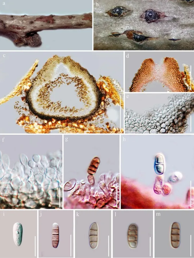
Saprobic or lemah patogen pada cabang mati dari Prunus armeniaca. Morph seksual tidak ditentukan. Morph asexual konidiomata 220–265 μm tinggi, 260–380 μm diam. (\(\bar{x} \) = 245 × 340 μm, n = 10), pycnidial, solitary, beredar, semi-mermermeredam di inang, subglobosa untuk ampulliform, ostiolate tidak jelas. Pycnidial walls 25–35 μm luas, lapisan luar dan interior terdiri dari sel cokelat gelap, 3–4 lapisan dan dinding tebal, hialin sel-sel tekstura angularis. Sel Conidiophores reduced ke konidiogen. Conidiogenous cells 10–22 μm × 4–6 μm, holoblastik, ampulliform, hialin, halus, bengkak di dasar, diskrit, menghasilkan satu konidium di puncak.
Profil ini diimpor dari uraian spesies pada sumber berlisensi dan sebagian teks diterjemahkan otomatis. Pemeriksaan editorial per profil belum selesai. Bagian yang belum diterjemahkan atau belum lolos pemeriksaan terjemahan tetap ditampilkan dalam bahasa sumber. Angka dan nama ilmiah dipertahankan.
Saprobic or weak pathogenic on dead twigs of Prunus armeniaca. Sexual morph Undetermined. Asexual morph Conidiomata 220–265 μm high, 260–380 μm diam. (\(\bar{x} \) = 245 × 340 μm, n = 10), pycnidial, solitary, scattered, semi-immersed in the host, subglobose to ampulliform, indistinct ostiolate. Pycnidial walls 25–35 μm wide, outer and inner layers composed of dark brown cells, 3–4 layers and thick-walled, hyaline cells of textura angularis. Conidiophores reduced to conidiogenous cells. Conidiogenous cells 10–22 μm × 4–6 μm, holoblastic, ampulliform, hyaline, smooth, swollen at the base, discrete, producing a single conidium at the apex. Conidia 11–16 × 6–10 μm (\(\bar{x} \) = 14 × 9 μm, n = 50), oblong to ellipsoidal, with rounded ends, occassionally truncate base, initially hyaline, unicellular, becoming dark brown before release from the pycnidia, 3–4-septate, not constricted at the septa, with thickenned, septal bands, slightly thick-walled.
Sebagian uraian ditampilkan dalam bahasa sumber; terjemahannya belum tersedia atau belum lolos pemeriksaan.
Pengukuran mengikuti objek, spesimen atau kultur, dan metode yang disebutkan sumber. Baca uraian asli untuk konteks lengkap.
02 / Kehidupan
Habitat dan peran ekologis
Saprobic or weak pathogenic on dead twigs of Prunus armeniaca. Sexual morph Undetermined. Asexual morph Conidiomata 220–265 μm high, 260–380 μm diam. (\(\bar{x} \) = 245 × 340 μm, n = 10), pycnidial, solitary, scattered, semi-immersed in the host, subglobose to ampulliform, indistinct ostiolate. Pycnidial walls 25–35 μm wide, outer and inner layers composed of dark brown cells, 3–4 layers and thick-walled, hyaline cells of textura angularis. Conidiophores reduced to conidiogenous cells. Conidiogenous cells 10–22 μm × 4–6 μm, holoblastic, ampulliform, hyaline, smooth, swollen at the base, discrete, producing a single conidium at the apex. Conidia 11–16 × 6–10 μm (\(\bar{x} \) = 14 × 9 μm, n = 50), oblong to ellipsoidal, with rounded ends, occassionally truncate base, initially hyaline, unicellular, becoming dark brown before release from the pycnidia, 3–4-septate, not constricted at the septa, with thickenned, septal bands, slightly thick-walled. Material examined: RUSSIA, Rostov region, Shakhty City, private garden, on dead and dying branches of Prunus armeniaca L. (Rosaceae), 11 May 2017, T.S. Bulgakov, T-1834 (MFLU 17-2136, holotype).
Sebagian uraian ditampilkan dalam bahasa sumber; terjemahannya belum tersedia atau belum lolos pemeriksaan.
Saprobic or weak pathogenic on dead twigs of Prunus armeniaca. Sexual morph Undetermined. Asexual morph Conidiomata 220–265 μm high, 260–380 μm diam. (\(\bar{x} \) = 245 × 340 μm, n = 10), pycnidial, solitary, scattered, semi-immersed in the host, subglobose to ampulliform, indistinct ostiolate. Pycnidial walls 25–35 μm wide, outer and inner layers composed of dark brown cells, 3–4 layers and thick-walled, hyaline cells of textura angularis. Conidiophores reduced to conidiogenous cells. Conidiogenous cells 10–22 μm × 4–6 μm, holoblastic, ampulliform, hyaline, smooth, swollen at the base, discrete, producing a single conidium at the apex. Conidia 11–16 × 6–10 μm (\(\bar{x} \) = 14 × 9 μm, n = 50), oblong to ellipsoidal, with rounded ends, occassionally truncate base, initially hyaline, unicellular, becoming dark brown before release from the pycnidia, 3–4-septate, not constricted at the septa, with thickenned, septal bands, slightly thick-walled.
Sebagian uraian ditampilkan dalam bahasa sumber; terjemahannya belum tersedia atau belum lolos pemeriksaan.
Nama diterima mengikuti Catalogue of Life Base2026-09-11; uraian mempertahankan konsep dan nama sumber aslinya.
Preserve original monograph treatment localities, hosts and specimen/substrate scope. Materials examined document specimens, not the full global range.
Impor dan terjemahan otomatis belum diperiksa editorial per profil. Struktur reproduktif yang disebutkan tidak membuktikan seluruh siklus hidup diketahui.
Pengukuran pada uraian mengikuti spesimen, kultur, dan metode sumber; angka tidak diperluas ke semua populasi.
Catatan distribusi dalam narasi mengikuti sumbernya. Dokumentasi spesimen atau foto mengikuti catatan sumber. Bukti wilayah Indonesia dan penilaian konservasi tidak disimpulkan dari gambar.
05 / Dokumentasi
Melihat dengan konteks
Uzbekistanica pruni (MFLU 17-2136, holotype). a, b Appearance of conidiomata on host. c Section of conidioma. d Ostiole. e Pycnidial wall. f – h Conidiogenous cells (g, h = stained with congo red). i – m Conidia (j, l = stained with congo red). Scale bars: c = 200 µm, d = 50 µm, e = 20 µm, f – h = 15 µm, i – m = 10 µm
Jenis
Pelat ilmiah
Kredit
Penulis dan sumber artikel: Hyde, Kevin D.; Dong, Yang; Phookamsak, Rungtiwa; Jeewon, Rajesh; Bhat, D. Jayarama; Jones, E. B. Gareth; Liu, Ning-Guo; Abeywickrama, Pranami D.; Mapook, Ausana; Wei, Deping; Perera, Rekhani H.; Manawasinghe, Ishara S.; Pem, Dhandevi; Bundhun, Digvijayini; Karunarathna, Anuruddha; Ekanayaka, Anusha H.; Bao, Dan-Feng; Li, Junfu; Samarakoon, Milan C.; Chaiwan, Napalai; Lin, Chuan-Gen; Phutthacharoen, Kunthida; Zhang, Sheng-Nan; Senanayake, Indunil C.; Goonasekara, Ishani D.; Thambugala, Kasun M.; Phukhamsakda, Chayanard; Tennakoon, Danushka S.; Jiang, Hong-Bo; Yang, Jing; Zeng, Ming; Huanraluek, Naruemon; Liu, Jian-Kui (Jack); Wijesinghe, Subodini N.; Tian, Qing; Tibpromma, Saowaluck; Brahmanage, Rashika S.; Boonmee, Saranyaphat; Huang, Shi-Ke; Thiyagaraja, Vinodhini; Lu, Yong-Zhong; Jayawardena, Ruvishika S.; Dong, Wei; Yang, Er-Fu; Singh, Sanjay K.; Singh, Shiv Mohan; Rana, Shiwali; Lad, Sneha S.; Anand, Garima; Devadatha, Bandarupalli; Niranjan, M.; Sarma, V. Venkateswara; Liimatainen, Kare; Aguirre-Hudson, Begoña; Niskanen, Tuula; Overall, Andy; Alvarenga, Renato Lúcio Mendes; Gibertoni, Tatiana Baptista; Pfliegler, Walter P.; Horváth, Enikő; Imre, Alexandra; Alves, Amanda Lucia; da Silva Santos, Ana Carla; Tiago, Patricia Vieira; Bulgakov, Timur S.; Wanasinghe, Dhanushaka N.; Bahkali, Ali H.; Doilom, Mingkwan; Elgorban, Abdallah M.; Maharachchikumbura, Sajeewa S. N.; Rajeshkumar, Kunhiraman C.; Haelewaters, Danny; Mortimer, Peter E.; Zhao, Qi; Lumyong, Saisamorn; Xu, Jianchu; Sheng, Jun
Saprobic or weak pathogenic on dead twigs of Prunus armeniaca.
Sexual morph Undetermined.
Asexual morph Conidiomata 220–265 μm high, 260–380 μm diam. (\(\bar{x} \) = 245 × 340 μm, n = 10), pycnidial, solitary, scattered, semi-immersed in the host, subglobose to ampulliform, indistinct ostiolate.
Pycnidial walls 25–35 μm wide, outer and inner layers composed of dark brown cells, 3–4 layers and thick-walled, hyaline cells of textura angularis.
Conidiophores reduced to conidiogenous cells.
Conidiogenous cells 10–22 μm × 4–6 μm, holoblastic, ampulliform, hyaline, smooth, swollen at the base, discrete, producing a single conidium at the apex.
Saprobic or weak pathogenic on dead twigs of Prunus armeniaca. Sexual morph Undetermined. Asexual morph Conidiomata 220–265 μm high, 260–380 μm diam. (\(\bar{x} \) = 245 × 340 μm, n = 10), pycnidial, solitary, scattered, semi-immersed in the host, subglobose to ampulliform, indistinct ostiolate. Pycnidial walls 25–35 μm wide, outer and inner layers composed of dark brown cells, 3–4 layers and thick-walled, hyaline cells of textura angularis. Conidiophores reduced to conidiogenous cells. Conidiogenous cells 10–22 μm × 4–6 μm, holoblastic, ampulliform, hyaline, smooth, swollen at the base, discrete, producing a single conidium at the apex. Conidia 11–16 × 6–10 μm (\(\bar{x} \) = 14 × 9 μm, n = 50), oblong to ellipsoidal, with rounded ends, occassionally truncate base, initially hyaline, unicellular, becoming dark brown before release from the pycnidia, 3–4-septate, not constricted at the septa, with thickenned, septal bands, slightly thick-walled.
Saprobic or weak pathogenic on dead twigs of Prunus armeniaca. Sexual morph Undetermined. Asexual morph Conidiomata 220–265 μm high, 260–380 μm diam. (\(\bar{x} \) = 245 × 340 μm, n = 10), pycnidial, solitary, scattered, semi-immersed in the host, subglobose to ampulliform, indistinct ostiolate. Pycnidial walls 25–35 μm wide, outer and inner layers composed of dark brown cells, 3–4 layers and thick-walled, hyaline cells of textura angularis. Conidiophores reduced to conidiogenous cells. Conidiogenous cells 10–22 μm × 4–6 μm, holoblastic, ampulliform, hyaline, smooth, swollen at the base, discrete, producing a single conidium at the apex. Conidia 11–16 × 6–10 μm (\(\bar{x} \) = 14 × 9 μm, n = 50), oblong to ellipsoidal, with rounded ends, occassionally truncate base, initially hyaline, unicellular, becoming dark brown before release from the pycnidia, 3–4-septate, not constricted at the septa, with thickenned, septal bands, slightly thick-walled.
Material examined: RUSSIA, Rostov region, Shakhty City, private garden, on dead and dying branches of Prunus armeniaca L. (Rosaceae), 11 May 2017, T.S. Bulgakov, T-1834 (MFLU 17-2136, holotype).
Saprobic or weak pathogenic on dead twigs of Prunus armeniaca.
Sexual morph Undetermined.
Asexual morph Conidiomata 220–265 μm high, 260–380 μm diam. (\(\bar{x} \) = 245 × 340 μm, n = 10), pycnidial, solitary, scattered, semi-immersed in the host, subglobose to ampulliform, indistinct ostiolate.
Pycnidial walls 25–35 μm wide, outer and inner layers composed of dark brown cells, 3–4 layers and thick-walled, hyaline cells of textura angularis.
Conidiophores reduced to conidiogenous cells.
Conidiogenous cells 10–22 μm × 4–6 μm, holoblastic, ampulliform, hyaline, smooth, swollen at the base, discrete, producing a single conidium at the apex.
Conidia 11–16 × 6–10 μm (\(\bar{x} \) = 14 × 9 μm, n = 50), oblong to ellipsoidal, with rounded ends, occassionally truncate base, initially hyaline, unicellular, becoming dark brown before release from the pycnidia, 3–4-septate, not constricted at the septa, with thickenned, septal bands, slightly thick-walled.
Status: impor sumber; pemeriksaan editorial per profil belum selesai. Untuk klaim biologis, gunakan rujukan pada bagian terkait dan periksa konteks sumber aslinya.
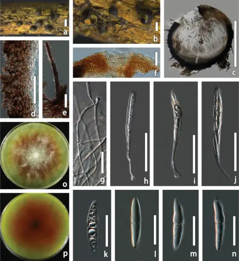
Saprobic on kayu yang membusuk dari Fagaceae. Morph seksual: Ascomata 586–690 × 425–630 μm (x̄ = 638 × 527 µm, n = 5), bercahaya, solitaris, sebagian besar dangkal, unilocular, globosa ke subglobosa, ditutupi dengan setae cokelat gelap…
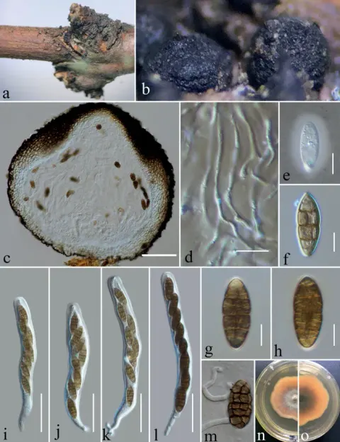
Saprobic on batang yang membusuk dari Rubus parvifolius dan Hypericum monogynum. Morph seksual: Ascomata 360–440 μm tinggi × 425–500 μm diam. (x̄ = 396 × 460 μm, n = 10), sebagian besar gregarious, hitam, globose ke subglobosa, dangkal,…
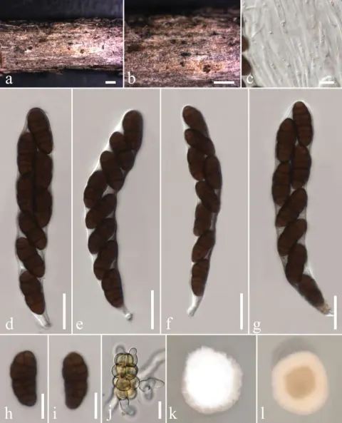
Saprobic on cabang mati dari Rosa ecae Aitch. Morph seksual: Ascomata 145–335 μm lebar, 70–195 μm tinggi, kesepian untuk gregarious, datar, semi-meremet ke permukaan, cokelat gelap ke hitam, globose untuk ovoid, karbonase, papilate. Peridi…