Ciri dan struktur
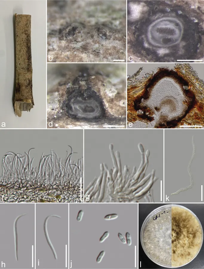
Saprobic on dead unidentified branch. Sexual morph: not observed. Asexual morph: Conidiomata 148–374 × 128–338 µm high (x̄ = 250 × 225 µm, n = 15), pycnidial, mostly scattered, immersed, slightly erumpent through the host surface, discoid or subglobose, with a solitary undivided locule. Conidiophores reduced to conidiogenous cells. Alpha conidiogenous cells 5.7–25 × 1.1–2.5 µm (x̄ = 15.6 × 1.7 µm, n = 50), hyaline, rarely branched, mostly aseptate, densely aggregated, cylindrical, straight to slightly curved and smooth. Alpha conidia 5–7.3 × 1.9–2.7 µm (x̄ = 6.3 × 2.3 µm, n = 40), unicellular, fusiform to ellipsoidal, apex and base rounded, hyaline, smooth, bi-guttulate. Beta conidiogenous cells 6.2–16 × 1.6–2.6 µm (x̄ = 9.4 × 2.1 µm, n = 40), phialidic, subcylindrical, tapering towards the apex, hyaline. Beta conidia 19–27 × 1–2 µm (x̄ = 23 × 1.5 µm, n = 40), filiform, aseptate, hyaline, smooth-walled, straight from base, and curve at apex. Gamma conidia not observed. Colonies on PDA, reaching 20 mm diam., after 3 weeks at 25 °C, initially white, turning beige after 7–10 days, flat, felty with a thick texture at the centre and marginal area, lacking aerial mycelium; reverse, glossy grey, radiating outwardly.
Habitat dan peran ekologis
Annona squamosa (Annonaceae), Berberis aristata (Berberidaceae), Carapa guianensis (Meliaceae), Citrus grandis cv. Tomentosa (Rutaceae), Cucumis melo (Cucurbitaceae), Glottidium sp. (Fabaceae), Glycine max, G. soja (Fabaceae), unidentified branch (Dong et al. 2021a, b; Hongsanan et al. 2023; This study). China, Myanmar, India, Indonesia, Japan, Thailand, the United States (Dong et al. 2021a, b; Hongsanan et al. 2023; this study).
- Habitat atau konteks temuan
- Informasi belum tersedia pada sumber rilis ini.
- Substrat
- Informasi belum tersedia pada sumber rilis ini.
- Peran atau konteks
- Belum dikategorikan pada rilis ini.
Reproduksi
Saprobic on dead unidentified branch. Sexual morph: not observed. Asexual morph: Conidiomata 148–374 × 128–338 µm high (x̄ = 250 × 225 µm, n = 15), pycnidial, mostly scattered, immersed, slightly erumpent through the host surface, discoid or subglobose, with a solitary undivided locule. Conidiophores reduced to conidiogenous cells. Alpha conidiogenous cells 5.7–25 × 1.1–2.5 µm (x̄ = 15.6 × 1.7 µm, n = 50), hyaline, rarely branched, mostly aseptate, densely aggregated, cylindrical, straight to slightly curved and smooth. Alpha conidia 5–7.3 × 1.9–2.7 µm (x̄ = 6.3 × 2.3 µm, n = 40), unicellular, fusiform to ellipsoidal, apex and base rounded, hyaline, smooth, bi-guttulate. Beta conidiogenous cells 6.2–16 × 1.6–2.6 µm (x̄ = 9.4 × 2.1 µm, n = 40), phialidic, subcylindrical, tapering towards the apex, hyaline.
Catatan identitas dan cakupan
- Nama diterima mengikuti Catalogue of Life Base2026-09-11; uraian mempertahankan konsep dan nama sumber aslinya.
- Preserve original treatment localities, hosts and substrate scope. Materials examined describe documented specimens, not the full global range.
- Impor dan terjemahan otomatis belum diperiksa editorial per profil. Struktur reproduktif yang disebutkan tidak membuktikan seluruh siklus hidup diketahui.
- Pengukuran pada uraian mengikuti spesimen, kultur, dan metode sumber; angka tidak diperluas ke semua populasi.
Catatan distribusi dalam narasi mengikuti sumbernya. Dokumentasi spesimen atau foto mengikuti catatan sumber. Bukti wilayah Indonesia dan penilaian konservasi tidak disimpulkan dari gambar.
Melihat dengan konteks
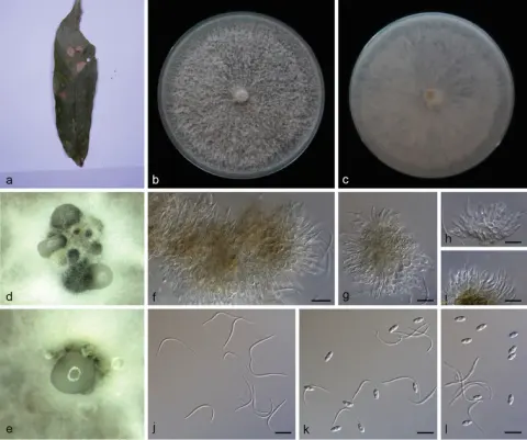
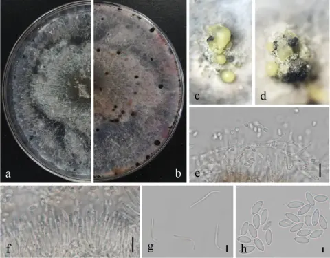
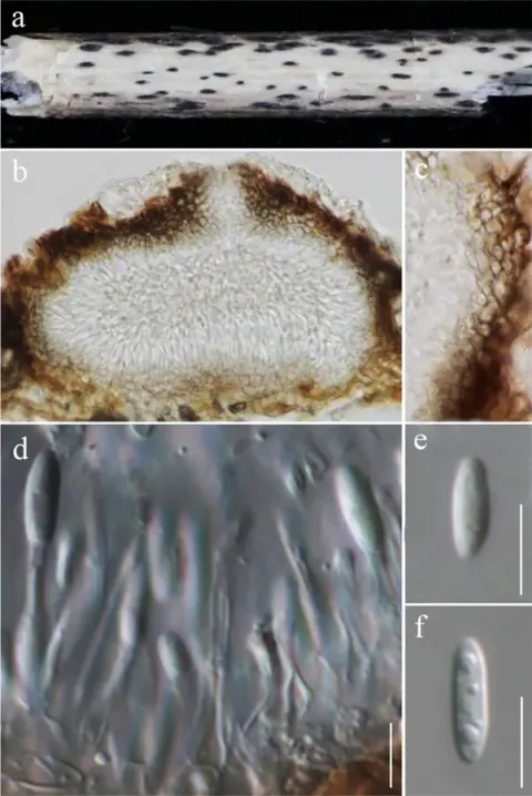
Diaporthe melonis (MFLU 23–0474) a host substrate b conidiomata on substrate c transverse section of conidioma d, e vertical section through conidiomata f, g conidiophores and conidiogenous cells h, i beta conidia j alpha conidia k a germinated conidium l front and reverse view of the colony on PDA. Scale bars: 200 μm (b–d); 100 μm (e); 20 μm (f, k); 10 μm (g, h–j).
- Jenis
- Pelat ilmiah
- Kredit
- Chayanard Phukhamsakda, Kevin D. Hyde, Milan C. Samarakoon, Johnny Louangphan, Kedsara Navasit, Fatimah Al-Otibi, Chitrabhanu S. Bhunjun
- Lisensi
- CC BY 4.0
- Sumber media
- Buka entri sumber
Sumber profil
- [1] Unveiling Sordariomycetes taxa associated with woody litter in Northern Thailand — Diaporthe melonis
- [2] Catalogue of Life Base2026-09-11 — Diaporthe melonis
- [3] Dokumentasi visual — Diaporthe melonis
Rujukan halaman ini
Fungi. Diaporthe melonis. Rilis data fungi-2026-10-12-expansion-2. https://fungi-atlas.mgadib.chatgpt.site/jamur/diaporthe-melonis/.
Status: impor sumber; pemeriksaan editorial per profil belum selesai. Untuk klaim biologis, gunakan rujukan pada bagian terkait dan periksa konteks sumber aslinya.