Ciri dan struktur
Saprobic on dead branches in a terrestrial habitat. Sexual morph: Undetermined. Asexual morph: Hyphomycetous. Colonies on natural substrate effuse, scattered, dark brown to black, hairy. Mycelium partly superficial, partly immersed in the substratum, composed of branched, septate, smooth, pale brown to brown hyphae. Conidiophores macronematous, mononematous, unbranched, solitary or in groups, erect, straight or slightly flexuous, cylindrical, smooth, brown to dark brown, 2–5-septate, robust at the base, 25–48.5 × 5–8 µm (x̄ = 34.9 × 6.1 µm, n = 12). Conidiogenous cells monoblastic, integrated, terminal, cylindrical, pale brown to brown, smooth, flat at the conidiogenous loco. Conidia acrogenous, solitary, dry, obclavate, straight or curved, reddish-brown and slightly paler towards the apex, 26–40-distoseptate, smooth, 150–270 × 11–15 µm (x̄ = 195.1 × 12 µm, n= 30), tapering to 2.5–7.5 µm near the apex, 2.5–5.5 µm wide at the truncate base. Colonies on PDA reaching 76–80 mm diam. after 4 weeks in an incubator under dark conditions at 25 °C, circular, surface velvety, with brown and denser mycelium at the center, becoming black at the margin with an obvious boundary, reverse black and sparser toward the boundary.
Habitat dan peran ekologis
Saprobic on dead branches in a terrestrial habitat. Sexual morph: Undetermined. Asexual morph: Hyphomycetous. Colonies on natural substrate effuse, scattered, dark brown to black, hairy. Mycelium partly superficial, partly immersed in the substratum, composed of branched, septate, smooth, pale brown to brown hyphae. Conidiophores macronematous, mononematous, unbranched, solitary or in groups, erect, straight or slightly flexuous, cylindrical, smooth, brown to dark brown, 2–5-septate, robust at the base, 25–48.5 × 5–8 µm (x̄ = 34.9 × 6.1 µm, n = 12). Conidiogenous cells monoblastic, integrated, terminal, cylindrical, pale brown to brown, smooth, flat at the conidiogenous loco. Conidia acrogenous, solitary, dry, obclavate, straight or curved, reddish-brown and slightly paler towards the apex, 26–40-distoseptate, smooth, 150–270 × 11–15 µm (x̄ = 195.1 × 12 µm, n= 30), tapering to 2.5–7.5 µm near the apex, 2.5–5.5 µm wide at the truncate base. Colonies on PDA reaching 76–80 mm diam. after 4 weeks in an incubator under dark conditions at 25 °C, circular, surface velvety, with brown and denser mycelium at the center, becoming black at the margin with an obvious boundary, reverse black and sparser toward the boundary.
- Habitat atau konteks temuan
- kultur yang didokumentasikan
- Substrat
- kayu
- Peran atau konteks
- Pengurai
Reproduksi
Saprobic on dead branches in a terrestrial habitat. Sexual morph: Undetermined. Asexual morph: Hyphomycetous. Colonies on natural substrate effuse, scattered, dark brown to black, hairy. Mycelium partly superficial, partly immersed in the substratum, composed of branched, septate, smooth, pale brown to brown hyphae. Conidiophores macronematous, mononematous, unbranched, solitary or in groups, erect, straight or slightly flexuous, cylindrical, smooth, brown to dark brown, 2–5-septate, robust at the base, 25–48.5 × 5–8 µm (x̄ = 34.9 × 6.1 µm, n = 12). Conidiogenous cells monoblastic, integrated, terminal, cylindrical, pale brown to brown, smooth, flat at the conidiogenous loco. Conidia acrogenous, solitary, dry, obclavate, straight or curved, reddish-brown and slightly paler towards the apex, 26–40-distoseptate, smooth, 150–270 × 11–15 µm (x̄ = 195.1 × 12 µm, n= 30), tapering to 2.5–7.5 µm near the apex, 2.5–5.5 µm wide at the truncate base.
Catatan identitas dan cakupan
- Nama diterima mengikuti Catalogue of Life Base2026-09-11; uraian mempertahankan konsep dan nama sumber aslinya.
- Preserve original treatment localities, hosts and substrate scope. Materials examined describe documented specimens, not the full global range.
- Impor dan terjemahan otomatis belum diperiksa editorial per profil. Struktur reproduktif yang disebutkan tidak membuktikan seluruh siklus hidup diketahui.
- Pengukuran pada uraian mengikuti spesimen, kultur, dan metode sumber; angka tidak diperluas ke semua populasi.
Catatan distribusi dalam narasi mengikuti sumbernya. Dokumentasi spesimen atau foto mengikuti catatan sumber. Bukti wilayah Indonesia dan penilaian konservasi tidak disimpulkan dari gambar.
Melihat dengan konteks
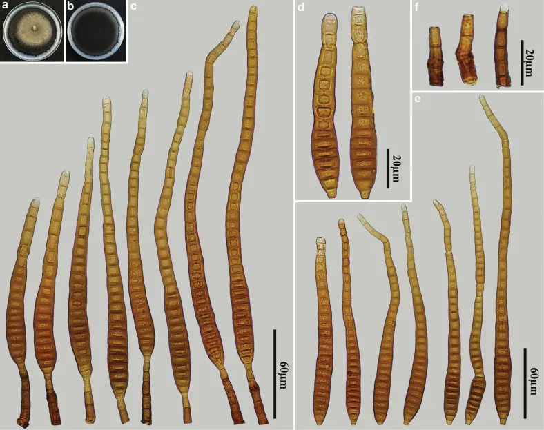
Distoseptispora clematidis (HJAUP M1319) a surface of colony after 4 weeks on PDA b reverse of colony after 4 weeks on PDA c conidiophores, conidiogenous cells and conidia d, e conidia f conidiophores.
- Jenis
- Pelat ilmiah
- Kredit
- Ming-Gen Liao, Xing-Xing Luo, Ya-Fen Hu, Rafael F. Castañeda-Ruíz, Zhao-Huan Xu, Jian Ma
- Lisensi
- CC BY 4.0
- Sumber media
- Buka entri sumber
Dokumentasi tambahan
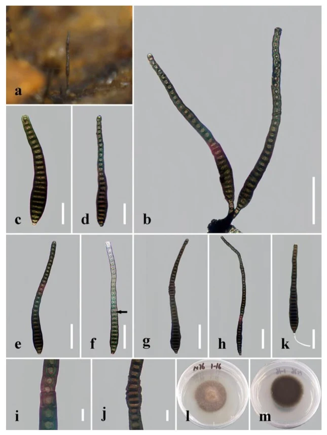
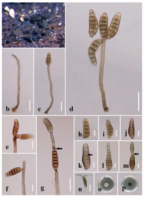
Distoseptispora clematidis. (a) Colonies on natural substrate. (b) Conidiophores with conidia. (c – h) Conidia. The arrow in f points to a gap in the middle of the septum, which indicates the distosepta. (i, j) Portions of conidia with percurrent proliferation in the middle of conidia. (k) Germinated conidium. (l) Colony on PDA (from front). (m) Colony on PDA (from reverse). Scale bars: (b, g – h) 50 μm; (c, d) 10 μm; (i, j) 5 μm.
Kredit dan penggunaan
© 2022 by the authors.
Ukuran dan format gambar disesuaikan tanpa pemotongan, retus, atau penambahan skala. Lisensi sumber dipertahankan.
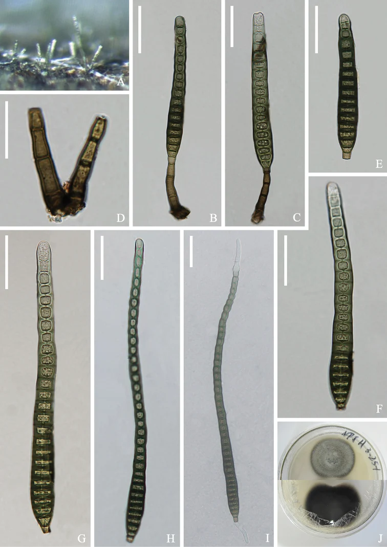
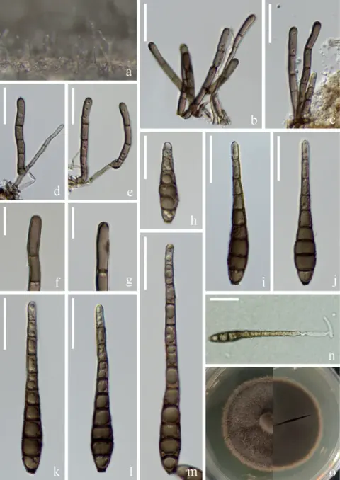
Distoseptispora clematidis (KUN-HKAS 112708) A colonies on the substratum B-C conidiophores with conidia D conidiogenous cells E-H conidia I germinating conidium J culture on PDA Scale bars: 30 μm (B, C, E-I); 20 μm (D).
Kredit dan penggunaan
Hong-Wei Shen, Dan-Feng Bao, Kevin D. Hyde, Hong-Yan Su, Darbhe J. Bhat, Zong-Long Luo
Ukuran dan format gambar disesuaikan tanpa pemotongan, retus, atau penambahan skala. Lisensi sumber dipertahankan.
Sumber profil
- [1] Morphological and phylogenetic analyses reveal four novel species of Distoseptispora (Distoseptisporaceae, Distoseptisporales) from southern China — Distoseptispora clematidis
- [2] Catalogue of Life Base2026-09-11 — Distoseptispora clematidis
- [3] Dokumentasi visual — Distoseptispora clematidis
- [4] Dokumentasi visual — Distoseptispora clematidis
- [5] Dokumentasi visual — Distoseptispora clematidis
Rujukan halaman ini
Fungi. Distoseptispora clematidis. Rilis data fungi-2026-10-12-expansion-2. https://fungi-atlas.mgadib.chatgpt.site/jamur/distoseptispora-clematidis/.
Status: impor sumber; pemeriksaan editorial per profil belum selesai. Untuk klaim biologis, gunakan rujukan pada bagian terkait dan periksa konteks sumber aslinya.