01 / Morfologi

Ciri dan struktur

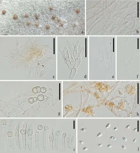
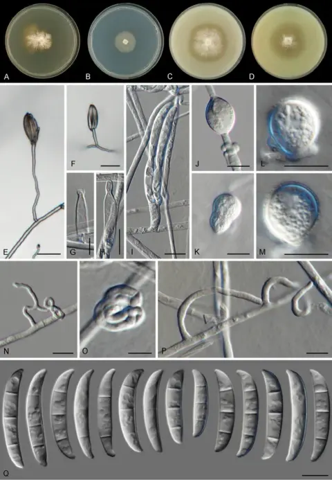
Sexual state not observed. Asexual state: Sporodochia not observed. Conidiophores borne on aerial mycelia, (8.6–)13.7–31.3(–50.3) μm, (mean ± SD = 22.5 ± 8.8 μm, n = 39), unbranched or branched, bearing terminal or intercalary monophialides, often reduced to single phialides; aerial phialides subulate to subcylindrical, smooth, thin-walled, (8.6–)10.9–19.5(–23.8) × (1.9–)2.5–3.8(–4.7) μm, (mean ± SD = 15.2 ± 4.3 × 3.2 ± 0.7 μm, n = 22), periclinal thickening inconspicuous or absent; aerial microconidia forming small false heads on tips of monophialides, hyaline, subglobose, oval, reniform or obovoid with a truncate base, ellipsoidal, smooth- and thin-walled, 0–1-septate, (6.3–)6.7–8.1(–8.7) × (1.9–)2–2.9(–3.2) μm, (mean ± SD = 7.4 ± 0.7 × 2.4 ± 0.5 μm, n = 33). Macroconidia and chlamydospores not observed. Colonies on PDA growing in the dark with an average growth rate of 10.3 mm/d at 25 °C. Colony colour white at first, becoming pale yellow, dense at the centre with radially sparse marginal mycelia. Aerial mycelium scant. Reverse pale yellow. Odour absent. Colonies on OMA incubated at 25 °C in the dark showing regular morphology, aerial mycelium sparse, covering the medium in five days. Reverse white without pigments. Colonies on SNA incubated at 25 °C in the dark exhibiting regular morphology, growing at 9.9 mm/d. Colony surface pure white, aerial mycelium scant. Reverse pure white, without diffusible pigments.

Sebagian uraian ditampilkan dalam bahasa sumber; terjemahannya belum tersedia atau belum lolos pemeriksaan.

Rujukan: [1]

Pengukuran mengikuti objek, spesimen atau kultur, dan metode yang disebutkan sumber. Baca uraian asli untuk konteks lengkap.

02 / Kehidupan

Habitat dan peran ekologis

From T. hybrid ‘Zhongshanshan’ in Jinan State-owned Beijiao Forest Farm, Jinan City, Shandong Province, China.

Sebagian uraian ditampilkan dalam bahasa sumber; terjemahannya belum tersedia atau belum lolos pemeriksaan.

Rujukan: [1]

Habitat atau konteks temuan
hutan
Substrat
Informasi belum tersedia pada sumber rilis ini.
Peran atau konteks
Belum dikategorikan pada rilis ini.
Temukan hubungan serupa
03 / Siklus hidup

Reproduksi

Sexual state not observed. Asexual state: Sporodochia not observed. Conidiophores borne on aerial mycelia, (8.6–)13.7–31.3(–50.3) μm, (mean ± SD = 22.5 ± 8.8 μm, n = 39), unbranched or branched, bearing terminal or intercalary monophialides, often reduced to single phialides; aerial phialides subulate to subcylindrical, smooth, thin-walled, (8.6–)10.9–19.5(–23.8) × (1.9–)2.5–3.8(–4.7) μm, (mean ± SD = 15.2 ± 4.3 × 3.2 ± 0.7 μm, n = 22), periclinal thickening inconspicuous or absent; aerial microconidia forming small false heads on tips of monophialides, hyaline, subglobose, oval, reniform or obovoid with a truncate base, ellipsoidal, smooth- and thin-walled, 0–1-septate, (6.3–)6.7–8.1(–8.7) × (1.9–)2–2.9(–3.2) μm, (mean ± SD = 7.4 ± 0.7 × 2.4 ± 0.5 μm, n = 33).

Sebagian uraian ditampilkan dalam bahasa sumber; terjemahannya belum tersedia atau belum lolos pemeriksaan.

Rujukan: [1]

04 / Batas informasi

Catatan identitas dan cakupan

  • Nama diterima mengikuti Catalogue of Life Base2026-09-11; uraian mempertahankan konsep dan nama sumber aslinya.
  • Preserve original treatment localities, hosts and substrate scope. Materials examined describe documented specimens, not the full global range.
  • Impor dan terjemahan otomatis belum diperiksa editorial per profil. Struktur reproduktif yang disebutkan tidak membuktikan seluruh siklus hidup diketahui.
  • Pengukuran pada uraian mengikuti spesimen, kultur, dan metode sumber; angka tidak diperluas ke semua populasi.

Catatan distribusi dalam narasi mengikuti sumbernya. Dokumentasi spesimen atau foto mengikuti catatan sumber. Bukti wilayah Indonesia dan penilaian konservasi tidak disimpulkan dari gambar.

05 / Dokumentasi

Melihat dengan konteks

Dokumentasi visual yang memenuhi pemeriksaan subjek dan hak penggunaan belum tersedia. Foto gejala, label, dan gambar yang meragukan tidak dihitung.

06 / Rujukan

Sumber profil

  1. [1] Two new species and four new host records of Fusarium species (Nectriaceae, Hypocreales) associated with Semanotus bifasciatus causing Taxodium hybrid ‘Zhongshanshan’ dieback — Fusarium annulatumBagian sumber: PMC13077318_section_13
    Tanggal penghimpunan sumber: 2026-10-12
    Teks sumber: CC BY 4.0 · He J, Li N, Li DW, Guo Y, Chen SS, Sun F, Wang JW, Huang L.
  2. [2] Catalogue of Life Base2026-09-11 — Fusarium annulatumBagian sumber: ColDP NameUsage.tsv ID 6JT4N; accepted species; source 2073
    Tanggal penghimpunan sumber: 2026-10-12

Adaptasi teks: CC BY 4.0. He J, Li N, Li DW, Guo Y, Chen SS, Sun F, Wang JW, Huang L. — Two new species and four new host records of Fusarium species (Nectriaceae, Hypocreales) associated with Semanotus bifasciatus causing Taxodium hybrid ‘Zhongshanshan’ dieback — Fusarium annulatum

Uraian asli dan riwayat sumber

Riwayat atau halaman sumber

Ringkasan sumber

Sexual state not observed. Asexual state: Sporodochia not observed. Conidiophores borne on aerial mycelia, (8.6–)13.7–31.3(–50.3) μm, (mean ± SD = 22.5 ± 8.8 μm, n = 39), unbranched or branched, bearing terminal or intercalary monophialides, often reduced to single phialides; aerial phialides subulate to subcylindrical, smooth, thin-walled, (8.6–)10.9–19.5(–23.8) × (1.9–)2.5–3.8(–4.7) μm, (mean ± SD = 15.2 ± 4.3 × 3.2 ± 0.7 μm, n = 22), periclinal thickening inconspicuous or absent; aerial microconidia forming small false heads on tips of monophialides, hyaline, subglobose, oval, reniform or obovoid with a truncate base, ellipsoidal, smooth- and thin-walled, 0–1-septate, (6.3–)6.7–8.1(–8.7) × (1.9–)2–2.9(–3.2) μm, (mean ± SD = 7.4 ± 0.7 × 2.4 ± 0.5 μm, n = 33).

Revisi sumber

Morfologi sumber

Sexual state not observed. Asexual state: Sporodochia not observed. Conidiophores borne on aerial mycelia, (8.6–)13.7–31.3(–50.3) μm, (mean ± SD = 22.5 ± 8.8 μm, n = 39), unbranched or branched, bearing terminal or intercalary monophialides, often reduced to single phialides; aerial phialides subulate to subcylindrical, smooth, thin-walled, (8.6–)10.9–19.5(–23.8) × (1.9–)2.5–3.8(–4.7) μm, (mean ± SD = 15.2 ± 4.3 × 3.2 ± 0.7 μm, n = 22), periclinal thickening inconspicuous or absent; aerial microconidia forming small false heads on tips of monophialides, hyaline, subglobose, oval, reniform or obovoid with a truncate base, ellipsoidal, smooth- and thin-walled, 0–1-septate, (6.3–)6.7–8.1(–8.7) × (1.9–)2–2.9(–3.2) μm, (mean ± SD = 7.4 ± 0.7 × 2.4 ± 0.5 μm, n = 33). Macroconidia and chlamydospores not observed. Colonies on PDA growing in the dark with an average growth rate of 10.3 mm/d at 25 °C. Colony colour white at first, becoming pale yellow, dense at the centre with radially sparse marginal mycelia. Aerial mycelium scant. Reverse pale yellow. Odour absent. Colonies on OMA incubated at 25 °C in the dark showing regular morphology, aerial mycelium sparse, covering the medium in five days. Reverse white without pigments. Colonies on SNA incubated at 25 °C in the dark exhibiting regular morphology, growing at 9.9 mm/d. Colony surface pure white, aerial mycelium scant. Reverse pure white, without diffusible pigments.

Revisi sumber

Ekologi sumber

From T. hybrid ‘Zhongshanshan’ in Jinan State-owned Beijiao Forest Farm, Jinan City, Shandong Province, China.

Revisi sumber

Struktur reproduksi sumber

Sexual state not observed. Asexual state: Sporodochia not observed. Conidiophores borne on aerial mycelia, (8.6–)13.7–31.3(–50.3) μm, (mean ± SD = 22.5 ± 8.8 μm, n = 39), unbranched or branched, bearing terminal or intercalary monophialides, often reduced to single phialides; aerial phialides subulate to subcylindrical, smooth, thin-walled, (8.6–)10.9–19.5(–23.8) × (1.9–)2.5–3.8(–4.7) μm, (mean ± SD = 15.2 ± 4.3 × 3.2 ± 0.7 μm, n = 22), periclinal thickening inconspicuous or absent; aerial microconidia forming small false heads on tips of monophialides, hyaline, subglobose, oval, reniform or obovoid with a truncate base, ellipsoidal, smooth- and thin-walled, 0–1-septate, (6.3–)6.7–8.1(–8.7) × (1.9–)2–2.9(–3.2) μm, (mean ± SD = 7.4 ± 0.7 × 2.4 ± 0.5 μm, n = 33).

Revisi sumber

Rujukan halaman ini

Fungi. Fusarium annulatum. Rilis data fungi-2026-10-12-expansion-2. https://fungi-atlas.mgadib.chatgpt.site/jamur/fusarium-annulatum/.

Status: impor sumber; pemeriksaan editorial per profil belum selesai. Untuk klaim biologis, gunakan rujukan pada bagian terkait dan periksa konteks sumber aslinya.