Ciri dan struktur
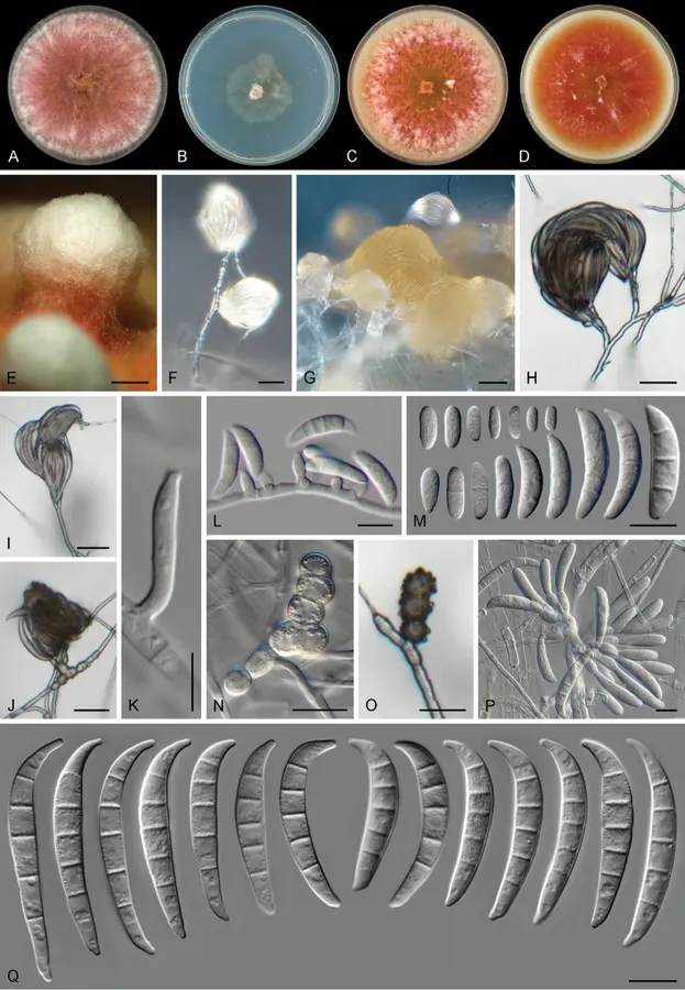
Different from most species in FSAMSC by its slender sporodochial conidia with tapered and somewhat rounded apex; its smooth- to tuberculate, often pigmented chlamydospores and the formation of large mycelial tufts on OA. Colonies on PDA growing in the dark with an average radial growth rate of 8.5–9.3 mm/d, reaching 34–37 mm diam. in 7 d at 24 °C, filling an entire 9 cm Petri dish in 7 d at 27–33 °C. Minimum temperature for growth 12 °C, maximum 36 °C, optimal 27–30 °C. Colony surface at first white, turning coral to dark vinaceous with white periphery and abundant yellow hyphae at the centre; flat, velvety to woolly, with abundant aerial mycelium and erect hyphal strings reaching several mm tall; colony margins regular and filiform. Reverse with yellow, coral or dark vinaceous patches, coral diffusible pigments strongly produced between 15–30 °C, turning scarlet to orange at 33–36 °C. Colonies on CMA and OA incubated at 24 °C in the dark occupying an entire 9 cm Petri dish in 7 d. Colony surface coral, rust to chestnut coloured in irregular patches, flat, felty to woolly, aerial mycelium scarce on CMA, mostly as radially dispersed white patches, on OA aerial mycelium abundant, especially on the periphery of the colony, forming dense, pustule-like, white mycelial tufts, formed by abundant intermingled hyphae and chlamydospores, 1–1.5 cm tall, with flesh to coral coloured stipes; margins on CMA and OA regular. Reverse pale luteous with red to coral periphery. Sporulation abundant from conidiophores formed on the aerial mycelium, at the agar level and from sporodochia. Conidiophores on the aerial mycelium straight or flexuous, septate, smooth- and thin-walled, up to 150 μm tall, sometimes emerging from irregular, swollen, pigmented and rough-walled cells on the hyphae; simple or sparingly and irregularly branched, branches bearing terminal, rarely lateral monophialides or reduced to conidiogenous cells borne laterally on hyphae; phialides on the aerial conidiophores short ampulliform, subulate to subcylindrical, smooth- and thin-walled, (7–)9–14(–15) μm long, (3–)4–5 μm at the widest point, without periclinal thickening and with a minute, inconspicuous collarette; conidia formed on aerial conidiophores of two types: a) hyaline, obovoid, ellipsoidal to clavate, smooth- and thin-walled, 0–1-septate, 2–14 × 2–4 μm; b) lunate to short falcate with a pointed apex and a somewhat flattened base, smooth- and thin-walled, 3–5-septate.
Habitat dan peran ekologis
Colonies on PDA growing in the dark with an average radial growth rate of 8.5–9.3 mm/d, reaching 34–37 mm diam. in 7 d at 24 °C, filling an entire 9 cm Petri dish in 7 d at 27–33 °C. Minimum temperature for growth 12 °C, maximum 36 °C, optimal 27–30 °C. Colony surface at first white, turning coral to dark vinaceous with white periphery and abundant yellow hyphae at the centre; flat, velvety to woolly, with abundant aerial mycelium and erect hyphal strings reaching several mm tall; colony margins regular and filiform. Reverse with yellow, coral or dark vinaceous patches, coral diffusible pigments strongly produced between 15–30 °C, turning scarlet to orange at 33–36 °C. Colonies on CMA and OA incubated at 24 °C in the dark occupying an entire 9 cm Petri dish in 7 d. Colony surface coral, rust to chestnut coloured in irregular patches, flat, felty to woolly, aerial mycelium scarce on CMA, mostly as radially dispersed white patches, on OA aerial mycelium abundant, especially on the periphery of the colony, forming dense, pustule-like, white mycelial tufts, formed by abundant intermingled hyphae and chlamydospores, 1–1.5 cm tall, with flesh to coral coloured stipes; margins on CMA and OA regular.
- Habitat atau konteks temuan
- kultur yang didokumentasikan
- Substrat
- Informasi belum tersedia pada sumber rilis ini.
- Peran atau konteks
- Belum dikategorikan pada rilis ini.
Reproduksi
Different from most species in FSAMSC by its slender sporodochial conidia with tapered and somewhat rounded apex; its smooth- to tuberculate, often pigmented chlamydospores and the formation of large mycelial tufts on OA. Colonies on PDA growing in the dark with an average radial growth rate of 8.5–9.3 mm/d, reaching 34–37 mm diam. in 7 d at 24 °C, filling an entire 9 cm Petri dish in 7 d at 27–33 °C. Minimum temperature for growth 12 °C, maximum 36 °C, optimal 27–30 °C. Colony surface at first white, turning coral to dark vinaceous with white periphery and abundant yellow hyphae at the centre; flat, velvety to woolly, with abundant aerial mycelium and erect hyphal strings reaching several mm tall; colony margins regular and filiform.
Catatan identitas dan cakupan
- Nama diterima mengikuti Catalogue of Life Base2026-09-11; uraian mempertahankan konsep dan nama sumber aslinya.
- Preserve original treatment localities, hosts and substrate scope. Materials examined describe documented specimens, not the full global range.
- Impor dan terjemahan otomatis belum diperiksa editorial per profil. Struktur reproduktif yang disebutkan tidak membuktikan seluruh siklus hidup diketahui.
- Pengukuran pada uraian mengikuti spesimen, kultur, dan metode sumber; angka tidak diperluas ke semua populasi.
Catatan distribusi dalam narasi mengikuti sumbernya. Dokumentasi spesimen atau foto mengikuti catatan sumber. Bukti wilayah Indonesia dan penilaian konservasi tidak disimpulkan dari gambar.
Melihat dengan konteks
Fusarium transvaalense sp. nov. A–D Colonies on PDA, SNA, OA and CMA, respectively, after 7 d at 24 °C in the dark E Pustule-like growth on OA F, G Sporodochia formed on the surface of carnation leaves H–L Aerial conidiophores phialides and conidia M Aerial conidia N, O Chlamydospores P Sporodochial conidiophores and phialides Q Sporodochial conidia. Scale bars: 2 mm (E); 20 μm (F–J); 5 μm (K); 10 μm (L–Q).
- Jenis
- Pelat ilmiah
- Kredit
- Marcelo Sandoval-Denis, Wijnand J. Swart, Pedro W. Crous
- Lisensi
- CC BY 4.0
- Sumber media
- Buka entri sumber
Sumber profil
- [1] New Fusarium species from the Kruger National Park, South Africa — Fusarium transvaalense
- [2] Catalogue of Life Base2026-09-11 — Fusarium transvaalense
- [3] Dokumentasi visual — Fusarium transvaalense
Rujukan halaman ini
Fungi. Fusarium transvaalense. Rilis data fungi-2026-10-12-expansion-2. https://fungi-atlas.mgadib.chatgpt.site/jamur/fusarium-transvaalense/.
Status: impor sumber; pemeriksaan editorial per profil belum selesai. Untuk klaim biologis, gunakan rujukan pada bagian terkait dan periksa konteks sumber aslinya.