01 / Morfologi

Ciri dan struktur

Saprobic on sugary exudates from insects feeding on the surface of living leaves of Maling bamboo. Thallus superficial, branched, septate, pale brown, darkly pigmented at the septa, forming distinct constructional rings. Asexual morph. Conidiomata, pycnidial, it originates from aggregated hyphae, with a bulbous base that consists of parallel hyphae and is straight to slightly flexuous. Conidiomata (273–378 μm, x̄ = 324 μm, n = 10) long, base (8–19 μm, x̄ = 15 μm, n = 10) wide, stalk light brown, conidiomata with cylindrical stalks expanding apically into a funnel-shaped cupula. Conidiogenous region resembling a cupula (45–61 × 30–47 μm, x̄ = 51 × 37 μm, n = 10), located in the cupulate apex of the conidiomata, brown, composed of cylindrical, thin-walled cells. Conidia (2.8–5.4 × 2.1–2.7 μm, x̄ = 3.9 × 2.4 μm, n = 30), hyaline, single-celled, ellipsoidal, smooth-walled, guttulate with 1–2 distinct refractive oil droplets, exuding in creamy masses from the ostiole. Sexual morph. Undetermined. Colonies black, loose, velvety, with sparse to moderate aerial mycelium and entire margins, reaching 23.91 mm in diam at 26 °C on PDA after 14 days in darkness.

Sebagian uraian ditampilkan dalam bahasa sumber; terjemahannya belum tersedia atau belum lolos pemeriksaan.

Rujukan: [1]

Pengukuran mengikuti objek, spesimen atau kultur, dan metode yang disebutkan sumber. Baca uraian asli untuk konteks lengkap.

02 / Kehidupan

Habitat dan peran ekologis

Saprobic on sugary exudates from insects feeding on the surface of living leaves of Maling bamboo. Thallus superficial, branched, septate, pale brown, darkly pigmented at the septa, forming distinct constructional rings. Asexual morph. Conidiomata, pycnidial, it originates from aggregated hyphae, with a bulbous base that consists of parallel hyphae and is straight to slightly flexuous. Conidiomata (273–378 μm, x̄ = 324 μm, n = 10) long, base (8–19 μm, x̄ = 15 μm, n = 10) wide, stalk light brown, conidiomata with cylindrical stalks expanding apically into a funnel-shaped cupula. Conidiogenous region resembling a cupula (45–61 × 30–47 μm, x̄ = 51 × 37 μm, n = 10), located in the cupulate apex of the conidiomata, brown, composed of cylindrical, thin-walled cells. Conidia (2.8–5.4 × 2.1–2.7 μm, x̄ = 3.9 × 2.4 μm, n = 30), hyaline, single-celled, ellipsoidal, smooth-walled, guttulate with 1–2 distinct refractive oil droplets, exuding in creamy masses from the ostiole. Sexual morph. Undetermined. Colonies black, loose, velvety, with sparse to moderate aerial mycelium and entire margins, reaching 23.91 mm in diam at 26 °C on PDA after 14 days in darkness. China • Guizhou Province, Chishui City, on living leaves of Maling bamboo (28°2'1"N, 105°44'41"E), 12 April 2023, Yuwei Liu, CS1 (IFRD 99018), living culture IFRDCC 19-1219.

Sebagian uraian ditampilkan dalam bahasa sumber; terjemahannya belum tersedia atau belum lolos pemeriksaan.

Rujukan: [1]

Habitat atau konteks temuan
kultur yang didokumentasikan
Substrat
Informasi belum tersedia pada sumber rilis ini.
Peran atau konteks
Pengurai; Pembentuk liken
Temukan hubungan serupa
03 / Siklus hidup

Reproduksi

Saprobic on sugary exudates from insects feeding on the surface of living leaves of Maling bamboo. Thallus superficial, branched, septate, pale brown, darkly pigmented at the septa, forming distinct constructional rings. Asexual morph. Conidiomata, pycnidial, it originates from aggregated hyphae, with a bulbous base that consists of parallel hyphae and is straight to slightly flexuous. Conidiomata (273–378 μm, x̄ = 324 μm, n = 10) long, base (8–19 μm, x̄ = 15 μm, n = 10) wide, stalk light brown, conidiomata with cylindrical stalks expanding apically into a funnel-shaped cupula. Conidiogenous region resembling a cupula (45–61 × 30–47 μm, x̄ = 51 × 37 μm, n = 10), located in the cupulate apex of the conidiomata, brown, composed of cylindrical, thin-walled cells.

Sebagian uraian ditampilkan dalam bahasa sumber; terjemahannya belum tersedia atau belum lolos pemeriksaan.

Rujukan: [1]

Sinonim menurut acuan taksonomi rilis: Caldariomyces fumago
04 / Batas informasi

Catatan identitas dan cakupan

  • Nama diterima mengikuti Catalogue of Life Base2026-09-11; uraian mempertahankan konsep dan nama sumber aslinya.
  • Preserve original treatment localities, hosts and substrate scope. Materials examined describe documented specimens, not the full global range.
  • Impor dan terjemahan otomatis belum diperiksa editorial per profil. Struktur reproduktif yang disebutkan tidak membuktikan seluruh siklus hidup diketahui.
  • Pengukuran pada uraian mengikuti spesimen, kultur, dan metode sumber; angka tidak diperluas ke semua populasi.

Catatan distribusi dalam narasi mengikuti sumbernya. Dokumentasi spesimen atau foto mengikuti catatan sumber. Bukti wilayah Indonesia dan penilaian konservasi tidak disimpulkan dari gambar.

05 / Dokumentasi

Melihat dengan konteks

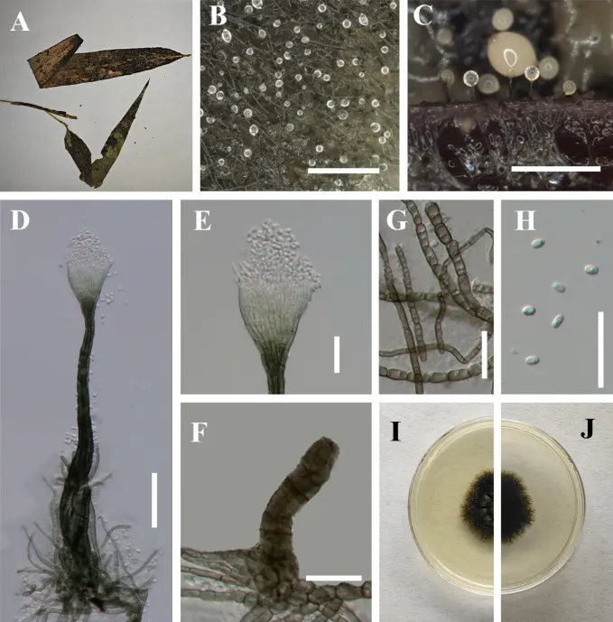
Leptoxyphium fumago (IFRDCC 19-1219). A Black mycelium covering the leaf surface; B, C Conidiomata on host; D Conidioma; E Cupulate ostiole; F Immature stages of conidioma; G Mycelial network; H Conidia; I Front view of the colony on PDA; J Reverse view of the colony on PDA. Scale bars: 1000 μm (B), 500 μm (C), 50 μm (D), 20 μm (E–H).

Jenis
Pelat ilmiah
Kredit
Jipeng Sun, Yuwei Liu, Tao Chen, Entaj Tarafder, Sinang Hongsanan, Shengxi Chen, Chada Norphanphoun, Xiaolu Deng, Yuzhe Feng, Tian Yang, Samantha C. Karunarathna, Xiangyu Zeng
Lisensi
CC BY 4.0
Sumber media
Buka entri sumber

Gambar menunjukkan objek menurut identifikasi sumber, bukan verifikasi independen spesimen oleh Fungi. Tidak ada skala baru yang ditambahkan.

06 / Rujukan

Sumber profil

  1. [1] Morphology and multilocus phylogeny illuminate novel taxa in Capnodiaceae (Ascomycota, Dothideomycetes, Capnodiales) — Leptoxyphium fumagoBagian sumber: PMC13241920_section_104
    Tanggal penghimpunan sumber: 2026-10-12
    Teks sumber: CC BY 4.0 · Sun J, Liu Y, Chen T, Tarafder E, Hongsanan S, Chen S, Norphanphoun C, Deng X, Feng Y, Yang T, Karunarathna SC, Zeng X.
  2. [2] Catalogue of Life Base2026-09-11 — Leptoxyphium fumagoBagian sumber: ColDP NameUsage.tsv ID 9Y2HG; accepted species; source 2073
    Tanggal penghimpunan sumber: 2026-10-12
  3. [3] Dokumentasi visual — Leptoxyphium fumagoBagian sumber: Individual media record; identification follows the source, not independent determination
    Tanggal penghimpunan sumber: 2026-10-12
    Media sumber: CC BY 4.0 · Jipeng Sun, Yuwei Liu, Tao Chen, Entaj Tarafder, Sinang Hongsanan, Shengxi Chen, Chada Norphanphoun, Xiaolu Deng, Yuzhe Feng, Tian Yang, Samantha C. Karunarathna, Xiangyu Zeng

Adaptasi teks: CC BY 4.0. Sun J, Liu Y, Chen T, Tarafder E, Hongsanan S, Chen S, Norphanphoun C, Deng X, Feng Y, Yang T, Karunarathna SC, Zeng X. — Morphology and multilocus phylogeny illuminate novel taxa in Capnodiaceae (Ascomycota, Dothideomycetes, Capnodiales) — Leptoxyphium fumago

Uraian asli dan riwayat sumber

Riwayat atau halaman sumber

Ringkasan sumber

Saprobic on sugary exudates from insects feeding on the surface of living leaves of Maling bamboo. Thallus superficial, branched, septate, pale brown, darkly pigmented at the septa, forming distinct constructional rings. Asexual morph. Conidiomata, pycnidial, it originates from aggregated hyphae, with a bulbous base that consists of parallel hyphae and is straight to slightly flexuous. Conidiomata (273–378 μm, x̄ = 324 μm, n = 10) long, base (8–19 μm, x̄ = 15 μm, n = 10) wide, stalk light brown, conidiomata with cylindrical stalks expanding apically into a funnel-shaped cupula.

Revisi sumber

Morfologi sumber

Saprobic on sugary exudates from insects feeding on the surface of living leaves of Maling bamboo. Thallus superficial, branched, septate, pale brown, darkly pigmented at the septa, forming distinct constructional rings. Asexual morph. Conidiomata, pycnidial, it originates from aggregated hyphae, with a bulbous base that consists of parallel hyphae and is straight to slightly flexuous. Conidiomata (273–378 μm, x̄ = 324 μm, n = 10) long, base (8–19 μm, x̄ = 15 μm, n = 10) wide, stalk light brown, conidiomata with cylindrical stalks expanding apically into a funnel-shaped cupula. Conidiogenous region resembling a cupula (45–61 × 30–47 μm, x̄ = 51 × 37 μm, n = 10), located in the cupulate apex of the conidiomata, brown, composed of cylindrical, thin-walled cells. Conidia (2.8–5.4 × 2.1–2.7 μm, x̄ = 3.9 × 2.4 μm, n = 30), hyaline, single-celled, ellipsoidal, smooth-walled, guttulate with 1–2 distinct refractive oil droplets, exuding in creamy masses from the ostiole. Sexual morph. Undetermined. Colonies black, loose, velvety, with sparse to moderate aerial mycelium and entire margins, reaching 23.91 mm in diam at 26 °C on PDA after 14 days in darkness.

Revisi sumber

Ekologi sumber

Saprobic on sugary exudates from insects feeding on the surface of living leaves of Maling bamboo. Thallus superficial, branched, septate, pale brown, darkly pigmented at the septa, forming distinct constructional rings. Asexual morph. Conidiomata, pycnidial, it originates from aggregated hyphae, with a bulbous base that consists of parallel hyphae and is straight to slightly flexuous. Conidiomata (273–378 μm, x̄ = 324 μm, n = 10) long, base (8–19 μm, x̄ = 15 μm, n = 10) wide, stalk light brown, conidiomata with cylindrical stalks expanding apically into a funnel-shaped cupula. Conidiogenous region resembling a cupula (45–61 × 30–47 μm, x̄ = 51 × 37 μm, n = 10), located in the cupulate apex of the conidiomata, brown, composed of cylindrical, thin-walled cells. Conidia (2.8–5.4 × 2.1–2.7 μm, x̄ = 3.9 × 2.4 μm, n = 30), hyaline, single-celled, ellipsoidal, smooth-walled, guttulate with 1–2 distinct refractive oil droplets, exuding in creamy masses from the ostiole. Sexual morph. Undetermined. Colonies black, loose, velvety, with sparse to moderate aerial mycelium and entire margins, reaching 23.91 mm in diam at 26 °C on PDA after 14 days in darkness. China • Guizhou Province, Chishui City, on living leaves of Maling bamboo (28°2'1"N, 105°44'41"E), 12 April 2023, Yuwei Liu, CS1 (IFRD 99018), living culture IFRDCC 19-1219.

Revisi sumber

Struktur reproduksi sumber

Saprobic on sugary exudates from insects feeding on the surface of living leaves of Maling bamboo. Thallus superficial, branched, septate, pale brown, darkly pigmented at the septa, forming distinct constructional rings. Asexual morph. Conidiomata, pycnidial, it originates from aggregated hyphae, with a bulbous base that consists of parallel hyphae and is straight to slightly flexuous. Conidiomata (273–378 μm, x̄ = 324 μm, n = 10) long, base (8–19 μm, x̄ = 15 μm, n = 10) wide, stalk light brown, conidiomata with cylindrical stalks expanding apically into a funnel-shaped cupula. Conidiogenous region resembling a cupula (45–61 × 30–47 μm, x̄ = 51 × 37 μm, n = 10), located in the cupulate apex of the conidiomata, brown, composed of cylindrical, thin-walled cells.

Revisi sumber

Rujukan halaman ini

Fungi. Leptoxyphium fumago. Rilis data fungi-2026-10-12-expansion-2. https://fungi-atlas.mgadib.chatgpt.site/jamur/leptoxyphium-fumago/.

Status: impor sumber; pemeriksaan editorial per profil belum selesai. Untuk klaim biologis, gunakan rujukan pada bagian terkait dan periksa konteks sumber aslinya.