Ciri dan struktur
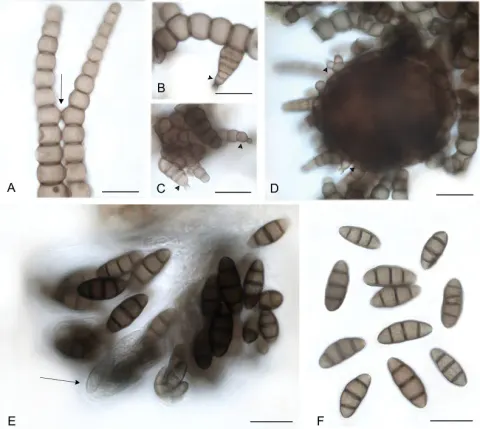
Subicula thick, dark brown to black, superficial. Mycelia of moniliform to doliiform hyphae, brown when mature, smooth to slightly verrucose throughout, and verrucose distally, tapered towards their ends. Cells broader than long, up to 30 µm wide, narrowing to 6 µm at hyphal tips. Sexual form. Ascomata scattered to crowded in some specimens, dark brown, semi–immersed in subicula, 120–200 × 90–150 µm, bearing moniliform appendages. No mature asci and ascospores observed. Asexual forms. Capnobotrys – No distinctive conidiophores observed. Conidiogenous cells arise variously, unilaterally from cells at and just below the hyphal tip, in clusters at the tips of erect hyphae. Conidiogenous cells ovoid, brown, verruculose, bearing denticulate scars where successive conidia budded out, then seceded. Conidia ovoid, walls up to 2 µm thick, up to five septate, slightly constricted at the septa, deeply verrucose, brown to dark brown. Conidium size increases with number of septa: aseptate conidia (6)–8.5–(10.5) +/– 1.7 × (5)–6.8–8) +/– 1.5 µm, N = 20, the two-septate conidia (17)–18.6–(20) +/– 1.7 × (8)–9–(11) +/– 1.0 µm, N = 26, the less common five-septate conidia (38.0)–41.0–(44.0) +/– 3.2 × (10.0)–11–(12.0) +/– 0.7 µm, N = 3. Cells wider than long, proximal cells usually wider and longer than distal cells. Capnophialophora – Phialides on distal cells of hyphae and (not illustrated) on capnobotrys conidia, light brown to brown, verruculose, subspherical venters 5–6 µm wide; collarettes deeply constricted at junction with venter, funnel–shaped, up to 4 µm long, opening to 2 µm wide. No phialidic conidia observed.
Habitat dan peran ekologis
Reported from New Zealand on living trunks of woody gymnosperms: Phyllocladus trichomanoides, Prumnopitys taxifolia, Dacrydium cupressinum (Hughes 1981). Possibly also widespread on Taxus baccata (Taxodiaceae) in Europe: specimens from Taxus sp. from Norway examined here, also reported from England (Ellis 1976), Ireland (Hughes 1981) and Italy (Karadelev et al. 2017).
- Habitat atau konteks temuan
- Informasi belum tersedia pada sumber rilis ini.
- Substrat
- Informasi belum tersedia pada sumber rilis ini.
- Peran atau konteks
- Belum dikategorikan pada rilis ini.
Reproduksi
Subicula thick, dark brown to black, superficial. Mycelia of moniliform to doliiform hyphae, brown when mature, smooth to slightly verrucose throughout, and verrucose distally, tapered towards their ends. Cells broader than long, up to 30 µm wide, narrowing to 6 µm at hyphal tips. Sexual form. Ascomata scattered to crowded in some specimens, dark brown, semi–immersed in subicula, 120–200 × 90–150 µm, bearing moniliform appendages. No mature asci and ascospores observed. Asexual forms. Capnobotrys – No distinctive conidiophores observed. Conidiogenous cells arise variously, unilaterally from cells at and just below the hyphal tip, in clusters at the tips of erect hyphae. Conidiogenous cells ovoid, brown, verruculose, bearing denticulate scars where successive conidia budded out, then seceded.
Catatan identitas dan cakupan
- Nama diterima mengikuti Catalogue of Life Base2026-09-11; uraian mempertahankan konsep dan nama sumber aslinya.
- Preserve original treatment localities, hosts and substrate scope. Materials examined describe documented specimens, not the full global range.
- Impor dan terjemahan otomatis belum diperiksa editorial per profil. Struktur reproduktif yang disebutkan tidak membuktikan seluruh siklus hidup diketahui.
- Pengukuran pada uraian mengikuti spesimen, kultur, dan metode sumber; angka tidak diperluas ke semua populasi.
Catatan distribusi dalam narasi mengikuti sumbernya. Dokumentasi spesimen atau foto mengikuti catatan sumber. Bukti wilayah Indonesia dan penilaian konservasi tidak disimpulkan dari gambar.
Melihat dengan konteks
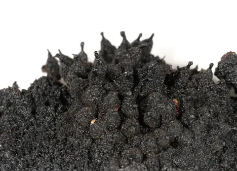
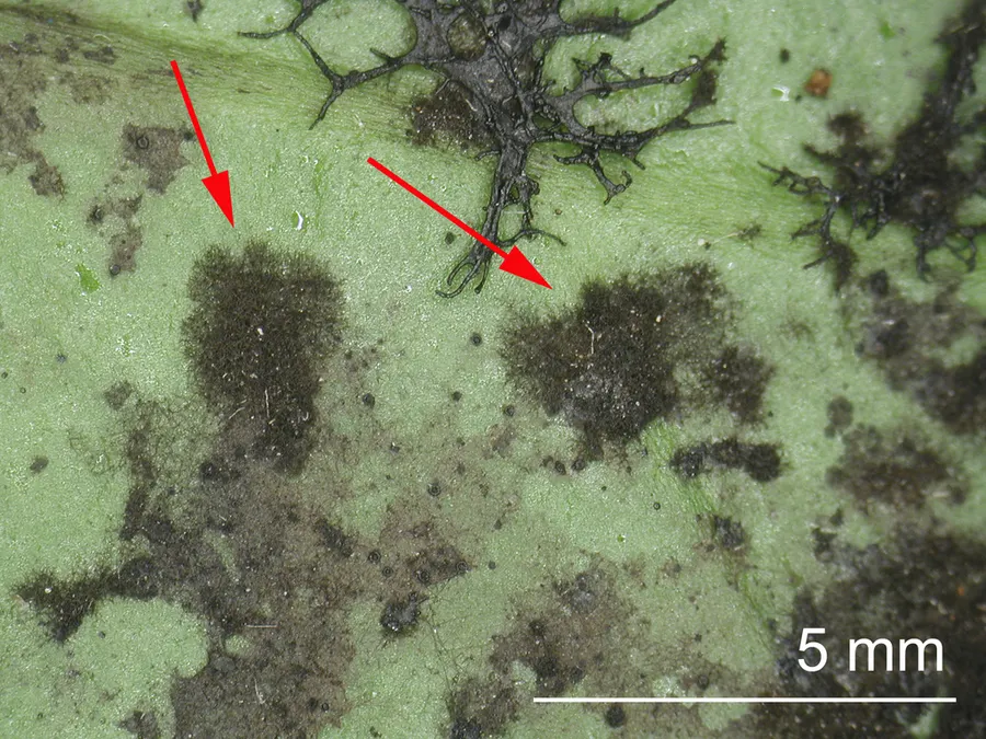
Dokumentasi Metacapnodium dingleyae menurut asosiasi foto pada record takson iNaturalist. Kredit: Jerry Cooper. Identitas mengikuti sumber; bukan penentuan spesimen independen.
- Jenis
- Fotografi
- Kredit
- (c) Jerry Cooper, some rights reserved (CC BY), uploaded by Jerry Cooper
- Lisensi
- CC BY 4.0
- Sumber media
- Buka entri sumber
Dokumentasi tambahan
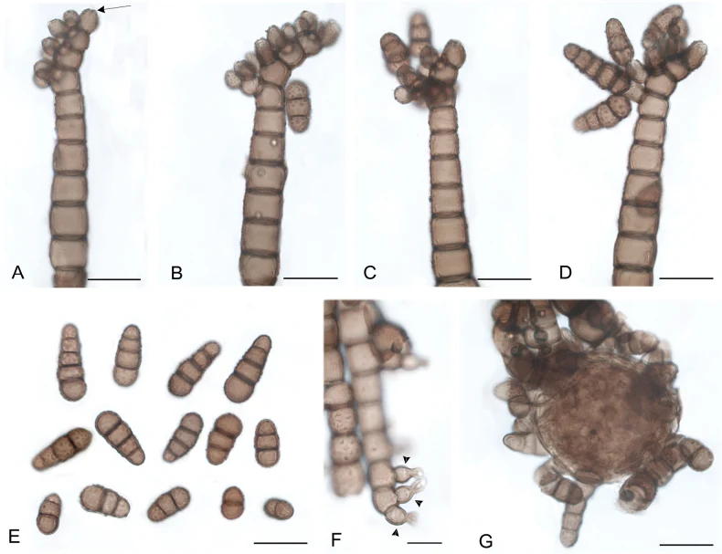
Metacapnodium dingleyae (O 201720). A–E. Capnobotrys form; A, B. Hyphae with conidiogenous cells; C, D. Initial and mature conidia; E. Conidia; F. Phialides (arrowheads); G. Ascoma. Scale bars: 20 μm (A–E, G); 10 μm (F).
Kredit dan penggunaan
Faezeh Aliabadi, Ludovic Le Renard, Mary L. Berbee
Ukuran dan format gambar disesuaikan tanpa pemotongan, retus, atau penambahan skala. Lisensi sumber dipertahankan.
Sumber profil
- [1] Taxonomy and phylogeny of the epiphytic sooty molds in family Metacapnodiaceae (class Eurotiomycetes, subclass Chaetothyriomycetidae) — Metacapnodium dingleyae
- [2] Catalogue of Life Base2026-09-11 — Metacapnodium dingleyae
- [3] Dokumentasi visual — Metacapnodium dingleyae
- [4] Dokumentasi visual — Metacapnodium dingleyae
Rujukan halaman ini
Fungi. Metacapnodium dingleyae. Rilis data fungi-2026-10-12-expansion-2. https://fungi-atlas.mgadib.chatgpt.site/jamur/metacapnodium-dingleyae/.
Status: impor sumber; pemeriksaan editorial per profil belum selesai. Untuk klaim biologis, gunakan rujukan pada bagian terkait dan periksa konteks sumber aslinya.