Ciri dan struktur
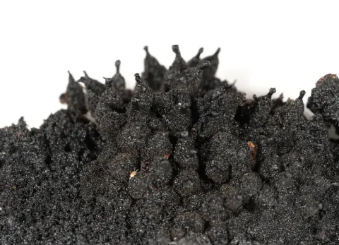
Subicula blackish, superficial, developing confluent thick and cracked cushions extending over host branches. Mycelia of doliiform and moniliform, brown to dark brown hyphae. Hyphae branching, straight, surface verrucose, tapering towards their tips. Hyphal cells to ~27 µm in diameter when mature, 5 µm in diameter at tip. Sexual form. Ascomata scattered to crowded, dark brown to black, globose, some subglobose, semi–immersed in subicula, 90–180(–200) × 90–180 µm, bearing moniliform appendages. No mature asci or ascospores observed. Asexual forms. Capnocybe – Synnemata up to 800 µm high, sparsely distributed throughout the subicula, their basal hyphae of ellipsoidal to globose cells, stipes 350–600 × 50–150 µm. Stipes of compact, fasciculate hyphae with cylindrical to ellipsoidal cells tapering to 6 µm towards the apex. Heads (capitula) of synnemata up to 300 µm wide. Terminal conidiogenous cells cylindrical, 10–16 × 5.5–7 µm, producing successive conidia blastically at their tips. Conidia fusiform, (2–) 3–septate, dark brown, (25)–33–(39) +/– 2.7 × (8)–9.2–(12) +/– 0.7 µm, Q = 3.6. Capnophialophora – Phialides light brown to brown, subspherical venters 5–6 µm wide, near-cylindrical collarettes up to 4 µm by 2–4 µm. No phialidic conidia observed.
Habitat dan peran ekologis
Hawksworth and Boluda (2020) reported the species from Spain and Portugal, commonly on Erica arborea, also on Erica australis, and perhaps present on other species of Erica.
- Habitat atau konteks temuan
- Informasi belum tersedia pada sumber rilis ini.
- Substrat
- Informasi belum tersedia pada sumber rilis ini.
- Peran atau konteks
- Belum dikategorikan pada rilis ini.
Reproduksi
Subicula blackish, superficial, developing confluent thick and cracked cushions extending over host branches. Mycelia of doliiform and moniliform, brown to dark brown hyphae. Hyphae branching, straight, surface verrucose, tapering towards their tips. Hyphal cells to ~27 µm in diameter when mature, 5 µm in diameter at tip. Sexual form. Ascomata scattered to crowded, dark brown to black, globose, some subglobose, semi–immersed in subicula, 90–180(–200) × 90–180 µm, bearing moniliform appendages. No mature asci or ascospores observed. Asexual forms. Capnocybe – Synnemata up to 800 µm high, sparsely distributed throughout the subicula, their basal hyphae of ellipsoidal to globose cells, stipes 350–600 × 50–150 µm.
Catatan identitas dan cakupan
- Nama diterima mengikuti Catalogue of Life Base2026-09-11; uraian mempertahankan konsep dan nama sumber aslinya.
- Preserve original treatment localities, hosts and substrate scope. Materials examined describe documented specimens, not the full global range.
- Impor dan terjemahan otomatis belum diperiksa editorial per profil. Struktur reproduktif yang disebutkan tidak membuktikan seluruh siklus hidup diketahui.
- Pengukuran pada uraian mengikuti spesimen, kultur, dan metode sumber; angka tidak diperluas ke semua populasi.
Catatan distribusi dalam narasi mengikuti sumbernya. Dokumentasi spesimen atau foto mengikuti catatan sumber. Bukti wilayah Indonesia dan penilaian konservasi tidak disimpulkan dari gambar.
Melihat dengan konteks
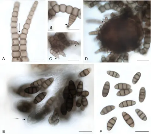
Sexual and asexual forms of Metacapnodiaceae. A. Phialides with a globose to subglobose venter (arrow) and a collarette at the neck, Metacapnodium ericophilum DAOM 234182; B. Globose to subglobose conidiogenous cells produce successive blastic conidia, M. stanhughesii UBC F35817; C. Synnema (arrow) of tightly compacted conidiophores, bearing conidiogenous cells and conidia at their tips, M. spongiosum (OSC61673); D. Capnosporium conidia with 2–4 septa, M. stanhughesii UBC F35817; E. Hormiokrypsis conidia with a straight, multicellular main axis and two multicellular, tapered arms, hormiokrypsis form of Ophiocapnocoma phloiophilia OSC61673; F. Ascoma with appendages, M. moniliforme DAOM 226251; G. Asci, M. adamantinum OSC 169460; H. Ascospores, M. moniliforme DAOM 226251. Scale bars: 5 µm (A); 20 μm (B, D–H); 50 µm (C).
- Jenis
- Pelat ilmiah
- Kredit
- Faezeh Aliabadi, Ludovic Le Renard, Mary L. Berbee
- Lisensi
- CC BY 4.0
- Sumber media
- Buka entri sumber
Dokumentasi tambahan
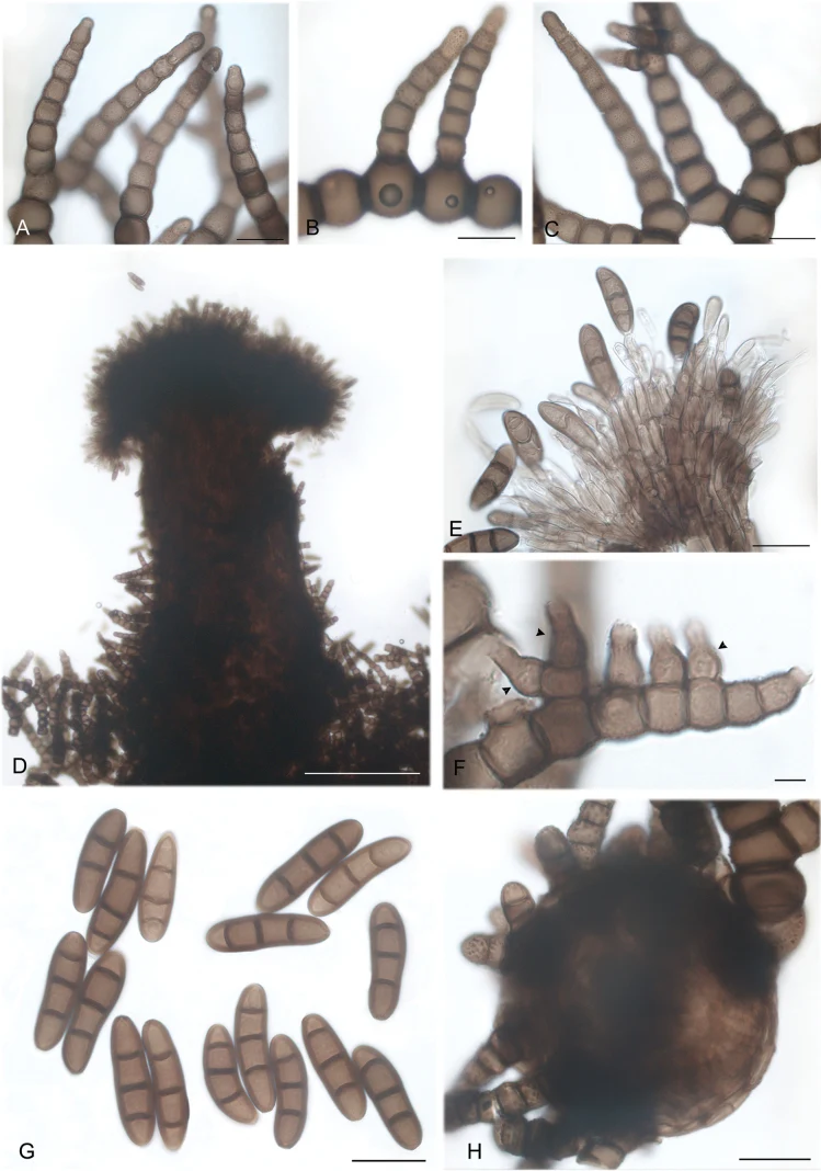
Metacapnodium ericophilum (DAOM 234182). A–C. Moniliform hyphae; D, E. Capnocybe form, synnema; D. Overview; E. Penicillate conidiogenous cells bearing conidia; F. Phialides with subcylindical collarettes (arrowheads); G. Conidia; H. Ascoma initial. Scale bars: 20 µm (A–C, E, G–H); 5 µm (F); 200 µm (D).
Kredit dan penggunaan
Faezeh Aliabadi, Ludovic Le Renard, Mary L. Berbee
Ukuran dan format gambar disesuaikan tanpa pemotongan, retus, atau penambahan skala. Lisensi sumber dipertahankan.
Sumber profil
- [1] Taxonomy and phylogeny of the epiphytic sooty molds in family Metacapnodiaceae (class Eurotiomycetes, subclass Chaetothyriomycetidae) — Metacapnodium ericophilum
- [2] Catalogue of Life Base2026-09-11 — Metacapnodium ericophilum
- [3] Dokumentasi visual — Metacapnodium ericophilum
- [4] Dokumentasi visual — Metacapnodium ericophilum
Rujukan halaman ini
Fungi. Metacapnodium ericophilum. Rilis data fungi-2026-10-12-expansion-2. https://fungi-atlas.mgadib.chatgpt.site/jamur/metacapnodium-ericophilum/.
Status: impor sumber; pemeriksaan editorial per profil belum selesai. Untuk klaim biologis, gunakan rujukan pada bagian terkait dan periksa konteks sumber aslinya.