Ciri dan struktur
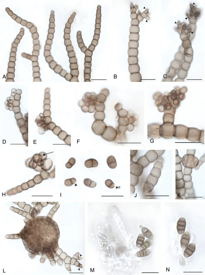
Subicula dark brown to black, effuse, superficial on leaves or twigs, typically less than 1 cm in diameter although sometimes becoming confluent. Mycelium of moniliform hyphae. Hyphae pale brown to brown, when mature, finely verrucose, deep constrictions between cells. Hyphae are erect, curved, smooth to finely verrucose, branching, then growing upwards, narrowing towards their tips. Most cells as broad as or broader than long, subglobose to doliiform, when mature up to 21 µm wide, tip cells narrowing to 5 µm. Sexual form. Ascomata scattered, semi-immersed in subicula, dark brown, globose (immature), subglobose to obovoid, (50–)70–180 µm high and 50–120 µm wide, ostiolate, having straight to curved appendages (Fig. 10K, L). Asci bitunicate, 8–spored, ellipsoid to clavate, 50–70 × 12–25 µm. Ascospores irregular or biseriate within asci, ellipsoid, light brown to brown, mainly 3(–4) septate, (17)–19.8–(23) +/– 2.6 × (8)–8.75–(10) +/– 0.7 µm (DAOM 226251). Asexual forms. Capnobotrys – No distinctive conidiophores observed. Conidiogenous cells unilateral or in botryose clusters at sides or tips of moniliform hyphae, globose to subglobose, brown to dark brown, bearing denticulate scars where successive conidia budded out, then seceded. Conidia brown, initially smooth walled, subglobose, elongating to become ellipsoid ovoid to allantoid, often cornute and inequilateral, with a single median or supramedial septum. Conidia (9)–14–(20) +/- 3.4 × (5)–7.7–(11) +/- 1.9 µm, Q = 1.8 (DAOM 226251) or (9)–12.3–(14.5) +/- 1.7 × (7)–7.9–(9) +/- 0.8 µm, Q = 1.6 (DAOM 234317, not illustrated).
Habitat dan peran ekologis
Reported on small twigs and leaves of varied plants including ferns and dicots. Widely distributed according to Hughes (1981); known from New Zealand, Australia, Chile, and perhaps elsewhere if M. cf. moniliforme UBC F33050 from Hawaii, and M. aff. moniliforme E 01043362 from Scotland could all be considered conspecific.
- Habitat atau konteks temuan
- Informasi belum tersedia pada sumber rilis ini.
- Substrat
- Informasi belum tersedia pada sumber rilis ini.
- Peran atau konteks
- Belum dikategorikan pada rilis ini.
Reproduksi
Subicula dark brown to black, effuse, superficial on leaves or twigs, typically less than 1 cm in diameter although sometimes becoming confluent. Mycelium of moniliform hyphae. Hyphae pale brown to brown, when mature, finely verrucose, deep constrictions between cells. Hyphae are erect, curved, smooth to finely verrucose, branching, then growing upwards, narrowing towards their tips. Most cells as broad as or broader than long, subglobose to doliiform, when mature up to 21 µm wide, tip cells narrowing to 5 µm. Sexual form. Ascomata scattered, semi-immersed in subicula, dark brown, globose (immature), subglobose to obovoid, (50–)70–180 µm high and 50–120 µm wide, ostiolate, having straight to curved appendages (Fig. 10K, L).
Catatan identitas dan cakupan
- Nama diterima mengikuti Catalogue of Life Base2026-09-11; uraian mempertahankan konsep dan nama sumber aslinya.
- Preserve original treatment localities, hosts and substrate scope. Materials examined describe documented specimens, not the full global range.
- Impor dan terjemahan otomatis belum diperiksa editorial per profil. Struktur reproduktif yang disebutkan tidak membuktikan seluruh siklus hidup diketahui.
- Pengukuran pada uraian mengikuti spesimen, kultur, dan metode sumber; angka tidak diperluas ke semua populasi.
Catatan distribusi dalam narasi mengikuti sumbernya. Dokumentasi spesimen atau foto mengikuti catatan sumber. Bukti wilayah Indonesia dan penilaian konservasi tidak disimpulkan dari gambar.
Melihat dengan konteks
Metacapnodium moniliforme (DAOM 226251). A. Moniliform hyphae; B, C. Moniliform hyphae with phialides (arrowheads); D–I. Capnobotrys form; D–G. Conidiogenous cells; H. Conidiogenous cell with denticles (arrow) from successive production and secession of conidia; I. Conidia, asterisks designate distal wall thinning; c designates a cornute conidium; J. Conidium of capnosporium form; K. Ascoma initial; L. Ascoma bearing phialides (arrowheads) on appendages; M. Immature and mature asci; N. Ascospores. Scale bars: 20 µm.
- Jenis
- Pelat ilmiah
- Kredit
- Faezeh Aliabadi, Ludovic Le Renard, Mary L. Berbee
- Lisensi
- CC BY 4.0
- Sumber media
- Buka entri sumber
Sumber profil
- [1] Taxonomy and phylogeny of the epiphytic sooty molds in family Metacapnodiaceae (class Eurotiomycetes, subclass Chaetothyriomycetidae) — Metacapnodium moniliforme
- [2] Catalogue of Life Base2026-09-11 — Metacapnodium moniliforme
- [3] Dokumentasi visual — Metacapnodium moniliforme
Rujukan halaman ini
Fungi. Metacapnodium moniliforme. Rilis data fungi-2026-10-12-expansion-2. https://fungi-atlas.mgadib.chatgpt.site/jamur/metacapnodium-moniliforme/.
Status: impor sumber; pemeriksaan editorial per profil belum selesai. Untuk klaim biologis, gunakan rujukan pada bagian terkait dan periksa konteks sumber aslinya.