Ciri dan struktur
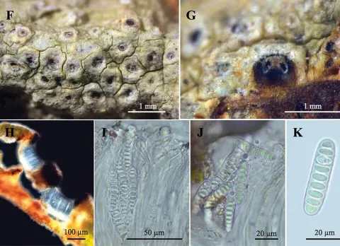
The lichen body (thallus) grows on bark and forms a continuous crust up to about 5 cm across. It is grayish and uneven, ranging from warty () to finely cracked (), with a white inner tissue (medulla) and no visible border zone (). In cross-section, the thallus is about 60–75 μm thick and contains scattered clusters of calcium oxalate crystals. The algal partner () is from the green algal genus Trentepohlia, with cells about 8–10 × 6–7 μm. The fruiting bodies (ascomata) are rounded and partly protruding from the thallus (), with a complete covering of thallus tissue (), measuring about 0.4–0.7 mm across and 0.15–0.2 mm high. The is hidden beneath a small pore-like opening about 0.07–0.1 mm wide, which is often partly filled by a central column () with a black tip (the columella may be sunken). The spore-bearing layer (hymenium) is colourless (hyaline) and about 120–150 μm high. Each ascus contains eight spores and measures about 100–110 × 12–15 μm. The ascospores are oval (ellipsoid), colourless, and divided into 8–9 cells (7–8-septate), measuring about 25–30 × 7.5–8 μm; they stain violet with iodine.
Habitat dan peran ekologis
The species is known from Pha Taem National Park in Ubon Ratchathani province, Thailand, where it was collected in dry evergreen forest at 124 m elevation, growing on bark near Sang Chan waterfall.
- Habitat atau konteks temuan
- hutan
- Substrat
- kulit kayu
- Peran atau konteks
- Pembentuk liken
Reproduksi
The fruiting bodies (ascomata) are rounded and partly protruding from the thallus (), with a complete covering of thallus tissue (), measuring about 0.4–0.7 mm across and 0.15–0.2 mm high. The is hidden beneath a small pore-like opening about 0.07–0.1 mm wide, which is often partly filled by a central column () with a black tip (the columella may be sunken). The spore-bearing layer (hymenium) is colourless (hyaline) and about 120–150 μm high. Each ascus contains eight spores and measures about 100–110 × 12–15 μm. The ascospores are oval (ellipsoid), colourless, and divided into 8–9 cells (7–8-septate), measuring about 25–30 × 7.5–8 μm; they stain violet with iodine.
Catatan identitas dan cakupan
- Nama diterima mengikuti Catalogue of Life Base2026-09-11; uraian mempertahankan konsep dan nama sumber aslinya.
- Original geographic, specimen and life-cycle scope retained; source import awaits individual editorial review.
- Impor dan terjemahan otomatis belum diperiksa editorial per profil. Struktur reproduktif yang disebutkan tidak membuktikan seluruh siklus hidup diketahui.
- Pengukuran pada uraian mengikuti spesimen, kultur, dan metode sumber; angka tidak diperluas ke semua populasi.
Catatan distribusi dalam narasi mengikuti sumbernya. Dokumentasi spesimen atau foto mengikuti catatan sumber. Bukti wilayah Indonesia dan penilaian konservasi tidak disimpulkan dari gambar.
Melihat dengan konteks
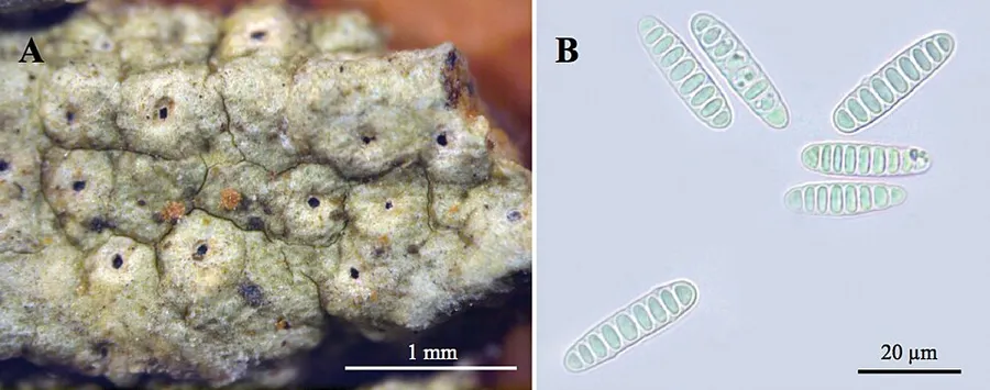
Dokumentasi Ocellularia phatamensis dari Wikimedia Commons. Figure 4A ; Morphology and anatomy of Ocellularia phatamensis (A–B) A habitat of ascomata, B ascospores (K. Papong 8574, holotype MSUT!). Scale bar A = 1 mm, B = 20 µm. Identifikasi mengikuti record berkas sumber.
- Jenis
- Fotografi
- Kredit
- Naksuwankul K, Kraichak E, Parnmen S, Lücking R,Lumbsch TH (2016) Five new species of Graphidaceae (Ascomycota, Ostropales) from Thailand. MycoKeys 17: 47-63. https://doi.org/10.3897/mycokeys.17.10512
- Lisensi
- CC BY 4.0
- Sumber media
- Buka entri sumber
Sumber profil
- [1] Ocellularia phatamensis — Wikipedia (en)
- [2] Catalogue of Life Base2026-09-11 — Ocellularia phatamensis
- [3] Dokumentasi visual — Ocellularia phatamensis
Rujukan halaman ini
Fungi. Ocellularia phatamensis. Rilis data fungi-2026-10-12-expansion-2. https://fungi-atlas.mgadib.chatgpt.site/jamur/ocellularia-phatamensis/.
Status: impor sumber; pemeriksaan editorial per profil belum selesai. Untuk klaim biologis, gunakan rujukan pada bagian terkait dan periksa konteks sumber aslinya.