Saprobic on daun rumput yang mati. Morph seksual tidak ditentukan. Transformasi aseksual Coelomycetous. konidiomata 140–290 µm tinggi, 135–240 µm berlian, piknidia, hitam, bercacah, gregaris, dangkal, satu-lokulasi, bervariasi dalam bentuk, subkonik hingga ovoid, atau subglobosa, dengan dasar truncate, paling lebar di dasar, glabrous, dengan ostiole tidak jelas. Conidiomata walls 17–100 µm lebar, tebal, tebal tidak sama, tebal di puncak, terdiri dari beberapa lapisan sel, lapisan luar yang terdiri dari sel-sel yang luas, cokelat gelap hingga hitam, sel-sel scleroplectenchymatous dari textura angularis hingga textura globulosa, lapisan dalam yang terdiri dari sel-sel yang luas, hialin textura angularis.
Profil ini diimpor dari uraian spesies pada sumber berlisensi dan sebagian teks diterjemahkan otomatis. Pemeriksaan editorial per profil belum selesai. Bagian yang belum diterjemahkan atau belum lolos pemeriksaan terjemahan tetap ditampilkan dalam bahasa sumber. Angka dan nama ilmiah dipertahankan.
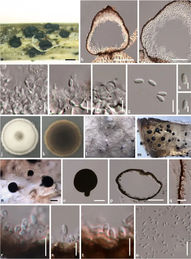
Plenodomus lijiangensis (KUN-HKAS 102249, holotype). a–i Morphological characteristics on natural substrate. l–u Morphological characteristics in vitro. a Conidiomata on host. b Vertical section of conidioma. c Section through conidioma wall. d–f Conidiogenous cells. g–i Conidia. j, k Culture characteristics on PDA (j = from above, k = from below). l–n Conidiomata forming on PDA after three months. o Squash mount of conidioma. p Vertical section of conidioma. q Section through conidioma wall stained with congo red. r, s Conidiogenous cells. t Conidiogenous cells stained with congo red. u Conidia. Scale bars a = 200 μm, b, n, o = 100 µm, c, p = 50 µm, u = 10 µm, g, q–t = 5 µm, h, i = 2 µm Penulis dan sumber artikel: Phookamsak, Rungtiwa; Hyde, Kevin D.; Jeewon, Rajesh; Bhat, D. Jayarama; Jones, E. B. Gareth; Maharachchikumbura, Sajeewa … (kredit lengkap di bawah) · CC BY 4.0 · Ukuran dan format gambar disesuaikan; lisensi asli dipertahankan.
01 / Morfologi
Ciri dan struktur
Saprobic on dead fronds of fern. Sexual morph Undetermined. Asexual morph Coelomycetous. Conidiomata 140–290 µm high, 135–240 µm diam., pycnidial, black, scattered, gregarious, superficial, uni-loculate, varied in shape, subconical to ovoid, or subglobose, with truncate base, widest at the base, glabrous, with indistinct ostiole. Conidiomata walls 17–100 µm wide, thick-walled, of unequal thickness, thickened at the apex, comprising several cell layers, outer layer composed of broad, dark brown to black, scleroplectenchymatous cells of textura angularis to textura globulosa, inner layer composed of broad, hyaline cells of textura angularis. Conidiophores reduced to conidiogenous cells. Conidiogenous cells 4–9 × 5–8 µm (\(\bar{x} \) = 6.4 × 6.5 μm, n = 40), enteroblastic, phialidic, discrete, determinate, ampulliform to doliiform, hyaline, smooth, with minute collarette, with 1–2 apertures, and periclinal wall thickening, arising from the inner cavity of the conidioma wall. Conidia 3–5 × 1.7–2.3 µm (\(\bar{x} \) = 4.3 × 2 µm, n = 50), hyaline, oblong to obovoid, aseptate, smooth-walled, with 1–2 guttules. Culture characteristics: Colonies on PDA, reaching 57–58 mm diam. after 3 weeks. Colony dense, circular, flattened, slightly raised, surface smooth, with edge entire, floccose; from above white at the margin, cream at the centre, with pale grey concentric ring near the margin; from below, yellowish brown at the edge, with paler margin, dark brown to black at the centre, colony slightly radiating outwards; not producing pigmentation on agar medium. Sporulation on PDA after three months.
Sebagian uraian ditampilkan dalam bahasa sumber; terjemahannya belum tersedia atau belum lolos pemeriksaan.
Pengukuran mengikuti objek, spesimen atau kultur, dan metode yang disebutkan sumber. Baca uraian asli untuk konteks lengkap.
02 / Kehidupan
Habitat dan peran ekologis
Saprobic on dead fronds of fern. Sexual morph Undetermined. Asexual morph Coelomycetous. Conidiomata 140–290 µm high, 135–240 µm diam., pycnidial, black, scattered, gregarious, superficial, uni-loculate, varied in shape, subconical to ovoid, or subglobose, with truncate base, widest at the base, glabrous, with indistinct ostiole. Conidiomata walls 17–100 µm wide, thick-walled, of unequal thickness, thickened at the apex, comprising several cell layers, outer layer composed of broad, dark brown to black, scleroplectenchymatous cells of textura angularis to textura globulosa, inner layer composed of broad, hyaline cells of textura angularis. Conidiophores reduced to conidiogenous cells. Conidiogenous cells 4–9 × 5–8 µm (\(\bar{x} \) = 6.4 × 6.5 μm, n = 40), enteroblastic, phialidic, discrete, determinate, ampulliform to doliiform, hyaline, smooth, with minute collarette, with 1–2 apertures, and periclinal wall thickening, arising from the inner cavity of the conidioma wall. Conidia 3–5 × 1.7–2.3 µm (\(\bar{x} \) = 4.3 × 2 µm, n = 50), hyaline, oblong to obovoid, aseptate, smooth-walled, with 1–2 guttules. Culture characteristics: Colonies on PDA, reaching 57–58 mm diam. after 3 weeks.
Sebagian uraian ditampilkan dalam bahasa sumber; terjemahannya belum tersedia atau belum lolos pemeriksaan.
Saprobic on dead fronds of fern. Sexual morph Undetermined. Asexual morph Coelomycetous. Conidiomata 140–290 µm high, 135–240 µm diam., pycnidial, black, scattered, gregarious, superficial, uni-loculate, varied in shape, subconical to ovoid, or subglobose, with truncate base, widest at the base, glabrous, with indistinct ostiole. Conidiomata walls 17–100 µm wide, thick-walled, of unequal thickness, thickened at the apex, comprising several cell layers, outer layer composed of broad, dark brown to black, scleroplectenchymatous cells of textura angularis to textura globulosa, inner layer composed of broad, hyaline cells of textura angularis. Conidiophores reduced to conidiogenous cells. Conidiogenous cells 4–9 × 5–8 µm (\(\bar{x} \) = 6.4 × 6.5 μm, n = 40), enteroblastic, phialidic, discrete, determinate, ampulliform to doliiform, hyaline, smooth, with minute collarette, with 1–2 apertures, and periclinal wall thickening, arising from the inner cavity of the conidioma wall.
Sebagian uraian ditampilkan dalam bahasa sumber; terjemahannya belum tersedia atau belum lolos pemeriksaan.
Nama diterima mengikuti Catalogue of Life Base2026-09-11; uraian mempertahankan konsep dan nama sumber aslinya.
Preserve original monograph treatment localities, hosts and specimen/substrate scope. Materials examined document specimens, not the full global range.
Impor dan terjemahan otomatis belum diperiksa editorial per profil. Struktur reproduktif yang disebutkan tidak membuktikan seluruh siklus hidup diketahui.
Pengukuran pada uraian mengikuti spesimen, kultur, dan metode sumber; angka tidak diperluas ke semua populasi.
Catatan distribusi dalam narasi mengikuti sumbernya. Dokumentasi spesimen atau foto mengikuti catatan sumber. Bukti wilayah Indonesia dan penilaian konservasi tidak disimpulkan dari gambar.
05 / Dokumentasi
Melihat dengan konteks
Plenodomus lijiangensis (KUN-HKAS 102249, holotype). a–i Morphological characteristics on natural substrate. l–u Morphological characteristics in vitro. a Conidiomata on host. b Vertical section of conidioma. c Section through conidioma wall. d–f Conidiogenous cells. g–i Conidia. j, k Culture characteristics on PDA (j = from above, k = from below). l–n Conidiomata forming on PDA after three months. o Squash mount of conidioma. p Vertical section of conidioma. q Section through conidioma wall stained with congo red. r, s Conidiogenous cells. t Conidiogenous cells stained with congo red. u Conidia. Scale bars a = 200 μm, b, n, o = 100 µm, c, p = 50 µm, u = 10 µm, g, q–t = 5 µm, h, i = 2 µm
Jenis
Pelat ilmiah
Kredit
Penulis dan sumber artikel: Phookamsak, Rungtiwa; Hyde, Kevin D.; Jeewon, Rajesh; Bhat, D. Jayarama; Jones, E. B. Gareth; Maharachchikumbura, Sajeewa S. N.; Raspé, Olivier; Karunarathna, Samantha C.; Wanasinghe, Dhanushka N.; Hongsanan, Sinang; Doilom, Mingkwan; Tennakoon, Danushka S.; Machado, Alexandre R.; Firmino, André L.; Ghosh, Aniket; Karunarathna, Anuruddha; Mešić, Armin; Dutta, Arun Kumar; Thongbai, Benjarong; Devadatha, Bandarupalli; Norphanphoun, Chada; Senwanna, Chanokned; Wei, Deping; Pem, Dhandevi; Ackah, Frank Kwekucher; Wang, Gen-Nuo; Jiang, Hong-Bo; Madrid, Hugo; Lee, Hyang Burm; Goonasekara, Ishani D.; Manawasinghe, Ishara S.; Kušan, Ivana; Cano, Josep; Gené, Josepa; Li, Junfu; Das, Kanad; Acharya, Krishnendu; Raj, K. N. Anil; Latha, K. P. Deepna; Chethana, K. W. Thilini; He, Mao-Qiang; Dueñas, Margarita; Jadan, Margita; Martín, María P.; Samarakoon, Milan C.; Dayarathne, Monika C.; Raza, Mubashar; Park, Myung Soo; Telleria, M. Teresa; Chaiwan, Napalai; Matočec, Neven; de Silva, Nimali I.; Pereira, Olinto L.; Singh, Paras Nath; Manimohan, Patinjareveettil; Uniyal, Priyanka; Shang, Qiu-Ju; Bhatt, Rajendra P.; Perera, Rekhani H.; Alvarenga, Renato Lúcio Mendes; Nogal-Prata, Sandra; Singh, Sanjay K.; Vadthanarat, Santhiti; Oh, Seung-Yoon; Huang, Shi-Ke; Rana, Shiwali; Konta, Sirinapa; Paloi, Soumitra; Jayasiri, Subashini C.; Jeon, Sun Jeong; Mehmood, Tahir; Gibertoni, Tatiana Baptista; Nguyen, Thuong T. T.; Singh, Upendra; Thiyagaraja, Vinodhini; Sarma, V. Venkateswara; Dong, Wei; Yu, Xian-Dong; Lu, Yong-Zhong; Lim, Young Woon; Chen, Yun; Tkalčec, Zdenko; Zhang, Zhi-Feng; Luo, Zong-Long; Daranagama, Dinushani A.; Thambugala, Kasun M.; Tibpromma, Saowaluck; Camporesi, Erio; Bulgakov, Timur S.; Dissanayake, Asha J.; Senanayake, Indunil C.; Dai, Dong Qin; Tang, Li-Zhou; Khan, Sehroon; Zhang, Huang; Promputtha, Itthayakorn; Cai, Lei; Chomnunti, Putarak; Zhao, Rui-Lin; Lumyong, Saisamorn; Boonmee, Saranyaphat; Wen, Ting-Chi; Mortimer, Peter E.; Xu, Jianchu
Saprobic on dead fronds of fern.
Sexual morph Undetermined.
Asexual morph Coelomycetous.
Conidiomata 140–290 µm high, 135–240 µm diam., pycnidial, black, scattered, gregarious, superficial, uni-loculate, varied in shape, subconical to ovoid, or subglobose, with truncate base, widest at the base, glabrous, with indistinct ostiole.
Conidiomata walls 17–100 µm wide, thick-walled, of unequal thickness, thickened at the apex, comprising several cell layers, outer layer composed of broad, dark brown to black, scleroplectenchymatous cells of textura angularis to textura globulosa, inner layer composed of broad, hyaline cells of textura angularis.
Saprobic on dead fronds of fern.
Sexual morph Undetermined.
Asexual morph Coelomycetous.
Conidiomata 140–290 µm high, 135–240 µm diam., pycnidial, black, scattered, gregarious, superficial, uni-loculate, varied in shape, subconical to ovoid, or subglobose, with truncate base, widest at the base, glabrous, with indistinct ostiole.
Conidiomata walls 17–100 µm wide, thick-walled, of unequal thickness, thickened at the apex, comprising several cell layers, outer layer composed of broad, dark brown to black, scleroplectenchymatous cells of textura angularis to textura globulosa, inner layer composed of broad, hyaline cells of textura angularis.
Conidiophores reduced to conidiogenous cells.
Conidiogenous cells 4–9 × 5–8 µm (\(\bar{x} \) = 6.4 × 6.5 μm, n = 40), enteroblastic, phialidic, discrete, determinate, ampulliform to doliiform, hyaline, smooth, with minute collarette, with 1–2 apertures, and periclinal wall thickening, arising from the inner cavity of the conidioma wall.
Conidia 3–5 × 1.7–2.3 µm (\(\bar{x} \) = 4.3 × 2 µm, n = 50), hyaline, oblong to obovoid, aseptate, smooth-walled, with 1–2 guttules.
Culture characteristics: Colonies on PDA, reaching 57–58 mm diam. after 3 weeks.
Colony dense, circular, flattened, slightly raised, surface smooth, with edge entire, floccose; from above white at the margin, cream at the centre, with pale grey concentric ring near the margin; from below, yellowish brown at the edge, with paler margin, dark brown to black at the centre, colony slightly radiating outwards; not producing pigmentation on agar medium.
Sporulation on PDA after three months.
Saprobic on dead fronds of fern.
Sexual morph Undetermined.
Asexual morph Coelomycetous.
Conidiomata 140–290 µm high, 135–240 µm diam., pycnidial, black, scattered, gregarious, superficial, uni-loculate, varied in shape, subconical to ovoid, or subglobose, with truncate base, widest at the base, glabrous, with indistinct ostiole.
Conidiomata walls 17–100 µm wide, thick-walled, of unequal thickness, thickened at the apex, comprising several cell layers, outer layer composed of broad, dark brown to black, scleroplectenchymatous cells of textura angularis to textura globulosa, inner layer composed of broad, hyaline cells of textura angularis.
Conidiophores reduced to conidiogenous cells.
Conidiogenous cells 4–9 × 5–8 µm (\(\bar{x} \) = 6.4 × 6.5 μm, n = 40), enteroblastic, phialidic, discrete, determinate, ampulliform to doliiform, hyaline, smooth, with minute collarette, with 1–2 apertures, and periclinal wall thickening, arising from the inner cavity of the conidioma wall.
Conidia 3–5 × 1.7–2.3 µm (\(\bar{x} \) = 4.3 × 2 µm, n = 50), hyaline, oblong to obovoid, aseptate, smooth-walled, with 1–2 guttules.
Culture characteristics: Colonies on PDA, reaching 57–58 mm diam. after 3 weeks.
Saprobic on dead fronds of fern.
Sexual morph Undetermined.
Asexual morph Coelomycetous.
Conidiomata 140–290 µm high, 135–240 µm diam., pycnidial, black, scattered, gregarious, superficial, uni-loculate, varied in shape, subconical to ovoid, or subglobose, with truncate base, widest at the base, glabrous, with indistinct ostiole.
Conidiomata walls 17–100 µm wide, thick-walled, of unequal thickness, thickened at the apex, comprising several cell layers, outer layer composed of broad, dark brown to black, scleroplectenchymatous cells of textura angularis to textura globulosa, inner layer composed of broad, hyaline cells of textura angularis.
Conidiophores reduced to conidiogenous cells.
Conidiogenous cells 4–9 × 5–8 µm (\(\bar{x} \) = 6.4 × 6.5 μm, n = 40), enteroblastic, phialidic, discrete, determinate, ampulliform to doliiform, hyaline, smooth, with minute collarette, with 1–2 apertures, and periclinal wall thickening, arising from the inner cavity of the conidioma wall.
Status: impor sumber; pemeriksaan editorial per profil belum selesai. Untuk klaim biologis, gunakan rujukan pada bagian terkait dan periksa konteks sumber aslinya.
Saprobic on daun kedelai mati dan batang mati dari Cirsium sp. Ascomata 250–290 µm tinggi, 300–360 µm berlian, hitam, bercahaya, bercahaya, gregarious, semi-merendam ke erumpent melalui epidermis inang, subglobosa ke subkonik,…
Saprobic on cabang dan batang mati dari Artemisia sp. Morph seksual Ascomata 140–280 μm tinggi, 300–450 μm berlian, hitam, bersinar di permukaan inang, tunggal hingga agregat, tenggelam di dasar, menjadi dangkal, uni-loculate, subglobosa…