Ciri dan struktur
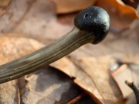
Growing on the stroma of Ophiocordyceps sp. on an Elateroidea larva. Stroma single, arising from the body end of the host larva, unbranched. The larva reddish-brown, cylindrical, 21 × 1.3–1.6 mm, intersegmental membranes conspicuous. Stipe of the stroma shiny black, stiff, band-like, but twisted and deeply wrinkled (dry specimen), more than 20 mm long and 1.0–1.3 mm thick, surface smooth (the fertile head was missing). Synnemata solitary or caespitose, arising from the intersegmental membranes of the larva and the surface of the stroma, mostly unbranched, generally straight, capitate, 1–3.5 mm long and 50–600 µm thick. Stipe basally broad and compressed, then gradually cylindrical upwards, white, greyish-white to yellowish-brown, surface smooth. Fertile head (including spore mass) abruptly expanded, ellipsoidal, 100–300 × 80–250 µm, located at the top of every synnema and distinctly separated from the stipe. Spore mass covers the surface of every fertile head, 15–25 µm thick, yellowish-brown and composed of hymenia. Phialides of two types, A-phialides produced on fertile heads, B-phialides arising laterally along the entire stipe. A-phialides 3–5 in terminal whorl on basal conidiophores, cylindrical to narrowly conical, straight or curved, non-uniform, 10–20 (x – = 15.1, n = 30) µm long and 1.5–2 µm (x – = 1.7, n = 30) wide, basally and terminally narrow, neck narrow to 0.5 µm, collarettes and periclinal thickening not visible; A-conidia obovate to obpyriform, smooth-walled, hyaline, 2.1–3.2 (x – = 2.6, n = 30) µm long and 1.5–2.2 (x – = 1.8, n = 30) µm wide. B-phialides single or in terminal whorls of 2–3 on basal conidiophores, straight, symmetrical or asymmetrical, hyaline, generally cylindrical, 10–25 (x – = 17, n = 30) µm long, 2–3.5 (x – = 2.8, n = 30) µm thick at the base, 0.5–0.8 (x – = 0.65, n =30) µm thick at the end, collarettes and periclinal thickening not visible; B-conidia fusiform, hyaline, smooth-walled, 3.2–6.0 (x – = 4.6, n = 30) µm long and 1–1.8 (x – = 1.4, n = 30) µm wide.
Habitat dan peran ekologis
On the stroma of Ophiocordyceps sp. on an Elateroidea larva on the ground in a humid bamboo (Chimonobambusa quadrangularis (Franceschi) Makino) forest in Guizhou karst regions. The larva might live in soil or decayed wood at first, but was then infected by Ophiocordyceps sp. and produced a sexual stroma. Following heavy rainfall, the host, together with the stroma of Ophiocordyceps sp., was washed away and exposed on the ground and at last, was parasitised by Polycephalomyces formosus. The fertile head of the stroma might have been lost during the floods.
- Habitat atau konteks temuan
- hutan
- Substrat
- tanah
- Peran atau konteks
- Belum dikategorikan pada rilis ini.
Reproduksi
Growing on the stroma of Ophiocordyceps sp. on an Elateroidea larva. Stroma single, arising from the body end of the host larva, unbranched. The larva reddish-brown, cylindrical, 21 × 1.3–1.6 mm, intersegmental membranes conspicuous. Stipe of the stroma shiny black, stiff, band-like, but twisted and deeply wrinkled (dry specimen), more than 20 mm long and 1.0–1.3 mm thick, surface smooth (the fertile head was missing). Synnemata solitary or caespitose, arising from the intersegmental membranes of the larva and the surface of the stroma, mostly unbranched, generally straight, capitate, 1–3.5 mm long and 50–600 µm thick. Stipe basally broad and compressed, then gradually cylindrical upwards, white, greyish-white to yellowish-brown, surface smooth.
Catatan identitas dan cakupan
- Nama diterima mengikuti Catalogue of Life Base2026-09-11; uraian mempertahankan konsep dan nama sumber aslinya.
- Preserve original treatment localities, hosts and substrate scope. Materials examined describe documented specimens, not the full global range.
- Impor dan terjemahan otomatis belum diperiksa editorial per profil. Struktur reproduktif yang disebutkan tidak membuktikan seluruh siklus hidup diketahui.
- Pengukuran pada uraian mengikuti spesimen, kultur, dan metode sumber; angka tidak diperluas ke semua populasi.
Catatan distribusi dalam narasi mengikuti sumbernya. Dokumentasi spesimen atau foto mengikuti catatan sumber. Bukti wilayah Indonesia dan penilaian konservasi tidak disimpulkan dari gambar.
Melihat dengan konteks
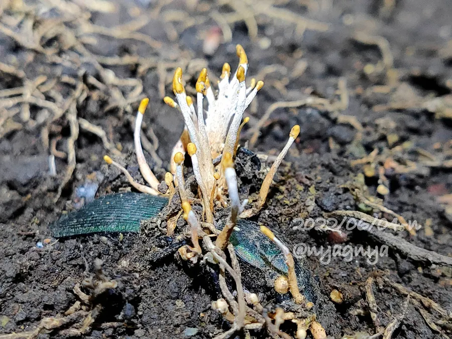
Dokumentasi Polycephalomyces formosus menurut asosiasi foto pada record takson iNaturalist. Kredit: Atsushi Nakajima. Identitas mengikuti sumber; bukan penentuan spesimen independen.
- Jenis
- Fotografi
- Kredit
- (c) Atsushi Nakajima, some rights reserved (CC BY), uploaded by Atsushi Nakajima
- Lisensi
- CC BY 4.0
- Sumber media
- Buka entri sumber
Dokumentasi tambahan
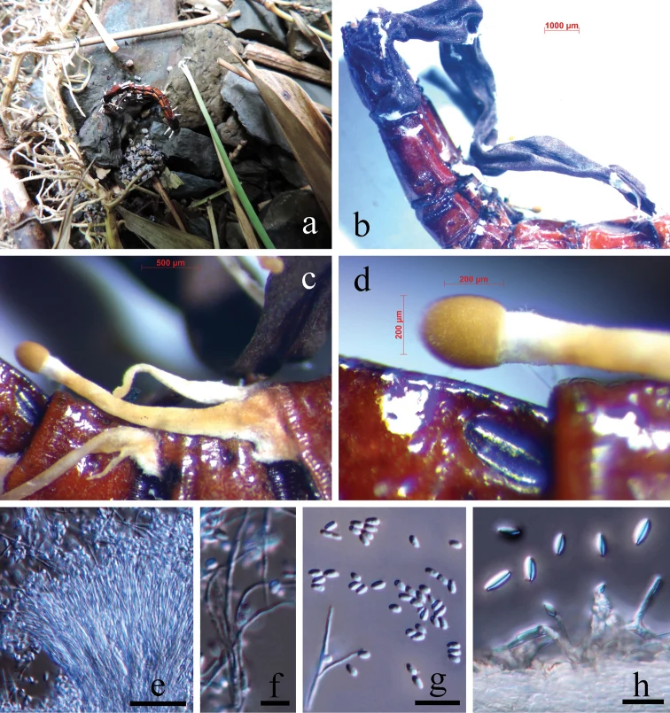
Polycephalomyces formosus (MFLU 18-0162) a collected on the ground in a bamboo forest b produced on the stroma of Ophiocordyceps sp. (the fertile head was missing) on an Elateroidea larva c, d synnemata e–g A-type phialides and A-type conidia h B-type phialides and B-type conidia. Scale bars: 20 µm (e); 5 µm (f); 10 µm (g, h).
Kredit dan penggunaan
Ling-Sheng Zha, Vadim Yu Kryukov, Jian-Hua Ding, Rajesh Jeewon, Putarak Chomnunti
Ukuran dan format gambar disesuaikan tanpa pemotongan, retus, atau penambahan skala. Lisensi sumber dipertahankan.
Sumber profil
- [1] Novel taxa and species diversity of Cordyceps sensu lato (Hypocreales, Ascomycota) developing on wireworms (Elateroidea and Tenebrionoidea, Coleoptera) — Polycephalomyces formosus
- [2] Catalogue of Life Base2026-09-11 — Polycephalomyces formosus
- [3] Dokumentasi visual — Polycephalomyces formosus
- [4] Dokumentasi visual — Polycephalomyces formosus
Rujukan halaman ini
Fungi. Polycephalomyces formosus. Rilis data fungi-2026-10-12-expansion-2. https://fungi-atlas.mgadib.chatgpt.site/jamur/polycephalomyces-formosus/.
Status: impor sumber; pemeriksaan editorial per profil belum selesai. Untuk klaim biologis, gunakan rujukan pada bagian terkait dan periksa konteks sumber aslinya.