01 / Morfologi

Ciri dan struktur

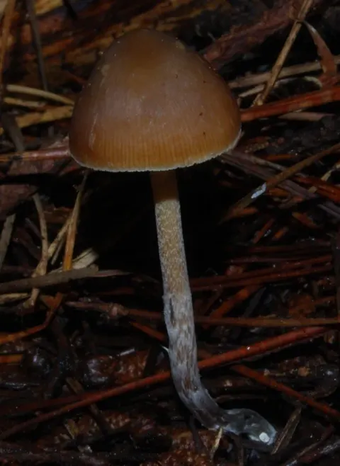
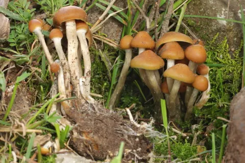
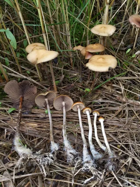
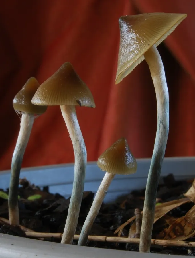
The cap is 10–40 mm in diameter, light brown to medium grey blue, conic to subcampanulate (cone-like to bell-shaped) with an inrolled margin and often an acute central papilla. It is translucent-striate to the margin (fine radial lines are visible around the edge of the cap when moist), extremely hygrophanous, glabrous (smooth or free of ornamentation) and slightly fibrous. The flesh inside is firm and brownish orange to yellowish. The gills are pale, thin and fairly close together, narrowly adnate (the gills meet the stipe by most of their width; they are broadly attached), with one or three short intermediate gills between two intermediate gills, and have a smooth edge. The stipe is 40–70 mm x 1–2 mm, pale greyish white, cylindrical, centered, fibrous, with brownish orange to yellowish flesh. It can be hollow or otherwise stuffed with fibres. The partial veil sometimes leaves a fragile line of raised threadlike tissue around the stipe close to halfway down. This can resemble a faint, thin raised ring, often stained blue. Spores measure 7.6–10.2(–11.5) × 5.8–8.1 × 4.7–7.1 μm. They are reddish grey, greyish orange to cinnamon brown in Meltzer's reagent, and appear subrhomboid in face view and ellipsoid to oval in side view. They are smooth with thick walls, and have a large eccentric germ pore which appears central in face view. This species has a very low spore production, often failing to produce a visible spore print. Basidia measure 20.9–27.2(–32.2) ×6.1–10.4 μm, are 4-spored, shaped broadly fusiform (like a spindle, rounded in the middle and tapering to the ends) to broadly clavate (shaped like a club).

Sebagian uraian ditampilkan dalam bahasa sumber; terjemahannya belum tersedia atau belum lolos pemeriksaan.

Rujukan: [1]

Pengukuran mengikuti objek, spesimen atau kultur, dan metode yang disebutkan sumber. Baca uraian asli untuk konteks lengkap.

02 / Kehidupan

Habitat dan peran ekologis

Scattered on heavily manured soil in grassland, and directly on cow dung, at Qingtiangang in Yangmingshan National Park in Taiwan. In potted plants and woodchip landscaping in New Zealand.

Sebagian uraian ditampilkan dalam bahasa sumber; terjemahannya belum tersedia atau belum lolos pemeriksaan.

Rujukan: [1]

Habitat atau konteks temuan
padang rumput
Substrat
Informasi belum tersedia pada sumber rilis ini.
Peran atau konteks
Belum dikategorikan pada rilis ini.
Temukan hubungan serupa
03 / Siklus hidup

Reproduksi

Spores measure 7.6–10.2(–11.5) × 5.8–8.1 × 4.7–7.1 μm. They are reddish grey, greyish orange to cinnamon brown in Meltzer's reagent, and appear subrhomboid in face view and ellipsoid to oval in side view. They are smooth with thick walls, and have a large eccentric germ pore which appears central in face view. This species has a very low spore production, often failing to produce a visible spore print. Basidia measure 20.9–27.2(–32.2) ×6.1–10.4 μm, are 4-spored, shaped broadly fusiform (like a spindle, rounded in the middle and tapering to the ends) to broadly clavate (shaped like a club). Pleurocystidia (the cystidia on the gill face) are absent or not well observed. Cheilocystidia (cystidia on the gill edge) measure 16.4–26.3(–29.2) μm long, (1.6–)1.8–3.0(–3.6) μm wide at the apex, (3.6–)4.5–7.1 μm wide at base.

Sebagian uraian ditampilkan dalam bahasa sumber; terjemahannya belum tersedia atau belum lolos pemeriksaan.

Rujukan: [1]

04 / Batas informasi

Catatan identitas dan cakupan

  • Nama diterima mengikuti Catalogue of Life Base2026-09-11; uraian mempertahankan konsep dan nama sumber aslinya.
  • Original geographic, specimen and life-cycle scope retained; source import awaits individual editorial review.
  • Impor dan terjemahan otomatis belum diperiksa editorial per profil. Struktur reproduktif yang disebutkan tidak membuktikan seluruh siklus hidup diketahui.
  • Pengukuran pada uraian mengikuti spesimen, kultur, dan metode sumber; angka tidak diperluas ke semua populasi.

Catatan distribusi dalam narasi mengikuti sumbernya. Dokumentasi spesimen atau foto mengikuti catatan sumber. Bukti wilayah Indonesia dan penilaian konservasi tidak disimpulkan dari gambar.

05 / Dokumentasi

Melihat dengan konteks

Dokumentasi Psilocybe angulospora menurut asosiasi foto pada record takson iNaturalist. Kredit: Alan Rockefeller. Identitas mengikuti sumber; bukan penentuan spesimen independen.

Jenis
Fotografi
Kredit
(c) Alan Rockefeller, some rights reserved (CC BY), uploaded by Alan Rockefeller
Lisensi
CC BY 4.0
Sumber media
Buka entri sumber

Gambar menunjukkan objek menurut identifikasi sumber, bukan verifikasi independen spesimen oleh Fungi. Tidak ada skala baru yang ditambahkan.

06 / Rujukan

Sumber profil

  1. [1] Psilocybe angulospora — Wikipedia (en)Bagian sumber: Frozen revision 1371341474
    Tanggal penghimpunan sumber: 2026-10-12
    Teks sumber: CC BY-SA4.0 · Wikipedia contributors
  2. [2] Catalogue of Life Base2026-09-11 — Psilocybe angulosporaBagian sumber: ColDP NameUsage.tsv ID 4PH2C; accepted species; source 2073
    Tanggal penghimpunan sumber: 2026-10-12
  3. [3] Dokumentasi visual — Psilocybe angulosporaBagian sumber: Individual media record; identification follows the source, not independent determination
    Tanggal penghimpunan sumber: 2026-10-12
    Media sumber: CC BY 4.0 · (c) Alan Rockefeller, some rights reserved (CC BY), uploaded by Alan Rockefeller

Adaptasi teks: CC BY-SA 4.0. Wikipedia contributors — Psilocybe angulospora — Wikipedia (en)

Uraian asli dan riwayat sumber

Riwayat atau halaman sumber

Ringkasan sumber

Psilocybe angulospora is a species of agaric fungus in the family Hymenogastraceae. The species was described from Taiwan in 2015 and is also present in New Zealand, where it is considered introduced. As a blueing member of the genus Psilocybe it contains the psychoactive compounds psilocin and psilocybin. The fruitbodies have a small, extremely hygrophanous pale gold conical to bell-shaped cap, often with a prominent pointed central papilla, a slender whitish stipe, and fine narrowly spaced gills.

Revisi sumber

Morfologi sumber

The cap is 10–40 mm in diameter, light brown to medium grey blue, conic to subcampanulate (cone-like to bell-shaped) with an inrolled margin and often an acute central papilla. It is translucent-striate to the margin (fine radial lines are visible around the edge of the cap when moist), extremely hygrophanous, glabrous (smooth or free of ornamentation) and slightly fibrous. The flesh inside is firm and brownish orange to yellowish. The gills are pale, thin and fairly close together, narrowly adnate (the gills meet the stipe by most of their width; they are broadly attached), with one or three short intermediate gills between two intermediate gills, and have a smooth edge. The stipe is 40–70 mm x 1–2 mm, pale greyish white, cylindrical, centered, fibrous, with brownish orange to yellowish flesh. It can be hollow or otherwise stuffed with fibres. The partial veil sometimes leaves a fragile line of raised threadlike tissue around the stipe close to halfway down. This can resemble a faint, thin raised ring, often stained blue. Spores measure 7.6–10.2(–11.5) × 5.8–8.1 × 4.7–7.1 μm. They are reddish grey, greyish orange to cinnamon brown in Meltzer's reagent, and appear subrhomboid in face view and ellipsoid to oval in side view. They are smooth with thick walls, and have a large eccentric germ pore which appears central in face view. This species has a very low spore production, often failing to produce a visible spore print. Basidia measure 20.9–27.2(–32.2) ×6.1–10.4 μm, are 4-spored, shaped broadly fusiform (like a spindle, rounded in the middle and tapering to the ends) to broadly clavate (shaped like a club).

Revisi sumber

Ekologi sumber

Scattered on heavily manured soil in grassland, and directly on cow dung, at Qingtiangang in Yangmingshan National Park in Taiwan. In potted plants and woodchip landscaping in New Zealand.

Revisi sumber

Struktur reproduksi sumber

Spores measure 7.6–10.2(–11.5) × 5.8–8.1 × 4.7–7.1 μm. They are reddish grey, greyish orange to cinnamon brown in Meltzer's reagent, and appear subrhomboid in face view and ellipsoid to oval in side view. They are smooth with thick walls, and have a large eccentric germ pore which appears central in face view. This species has a very low spore production, often failing to produce a visible spore print. Basidia measure 20.9–27.2(–32.2) ×6.1–10.4 μm, are 4-spored, shaped broadly fusiform (like a spindle, rounded in the middle and tapering to the ends) to broadly clavate (shaped like a club). Pleurocystidia (the cystidia on the gill face) are absent or not well observed. Cheilocystidia (cystidia on the gill edge) measure 16.4–26.3(–29.2) μm long, (1.6–)1.8–3.0(–3.6) μm wide at the apex, (3.6–)4.5–7.1 μm wide at base.

Revisi sumber

Rujukan halaman ini

Fungi. Psilocybe angulospora. Rilis data fungi-2026-10-12-expansion-2. https://fungi-atlas.mgadib.chatgpt.site/jamur/psilocybe-angulospora/.

Status: impor sumber; pemeriksaan editorial per profil belum selesai. Untuk klaim biologis, gunakan rujukan pada bagian terkait dan periksa konteks sumber aslinya.