Ciri dan struktur
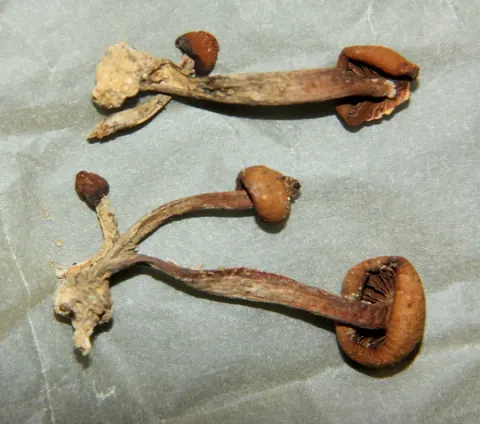
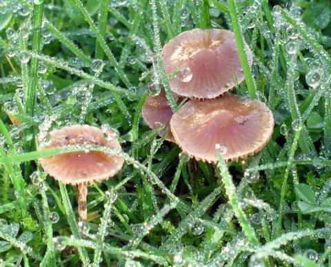
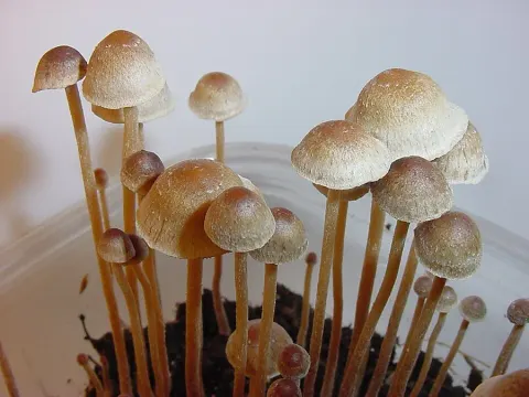
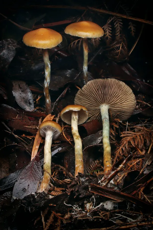
The cap averages 12–50 mm in diameter. It is conical to conic-convex with slightly inrolled edges when young, and becomes convex, often slightly upturned, sub-gibbous, or sometimes with a small acute umbo. It is slightly tacky. There may be veil remnants at the margin when young. Coloured yellow-brown to orange-brown, paler towards the margin, which is a little striate, hygrophanous, fading in drying to pallid biscuit brown or pale orange-yellow. The cap stains greenish blue with age or handling and the flesh inside is whitish. The gills are moderately close, pale smoky brown when young, violet-brown or brownish fuscous in age, with narrow pale edges, slightly ventricose, in three series, the middle reaching halfway to the stipe, with an adnate or broadly adnexed attachment and lines sometimes running down the stipe. The stipe is 25–70 × 2–3.5(-5) mm, tall and slender, equal or slightly wider towards the cap, finely vertically lined, mealy at the top with fine fibrils below, the base somewhat swollen or becoming a mass of mycelium, hollow inside, cartilaginous, pale whitish streaked with dark greyish brown, staining greenish blue, flesh brownish. A white cortinate partial veil soon disappears, leaving traces as a raised area around the upper stipe. Taste and odour are farinaceous and the spore print is purple brown. The cheilocystidia are 17–29 x 5.5–11 μm, hyaline, fusoid-ventricose, subpyriform or mucronate, often with an elongated neck at the apex which is 2–4.5 μm. The pleurocystidia measure 22–47 x 6–16 μm and is shaped like the cheilocystidia and also hyaline. The spores are smooth, subellipsoid, with an apical germ pore, measuring (10) 13.2–14.3 (15.4) x 6.6–7.7 x 6–7.5 μm.
Habitat dan peran ekologis
Psilocybe subaeruginosa grows solitary to gregarious from wood debris in Australian native forests, pine plantations and is occasionally observed growing on dung. Hal ini umum di bagian selatan Australia from April to Agustus. Spesies ini juga dikenal dari Australian native dan Eucalyptus forests, dan terkenal di New Zealand on kayu chip. Hal ini tercatat sebagai hadir di Selandia Baru, tetapi urutan DNA koleksi sejauh ini menunjukkan P. allenii dan P. cyanescens di sana serta P. subaeruginosa. Kecenderungan hidup dari kayu, serta dari tanaman dan tanah, berkontribusi pada kemungkinan bahwa ini adalah bahan yang ditanamnya ke Utara Hemisphere on, dan transplantasi menyebabkan penurunan keragaman genetik. Konsep ini juga menyebabkan gagasan bahwa mereka ada di daerah yang terganggu, bukan alami.
- Habitat atau konteks temuan
- hutan
- Substrat
- kayu; tanah; kotoran hewan
- Peran atau konteks
- Belum dikategorikan pada rilis ini.
Reproduksi
Taste and odour are farinaceous and the spore print is purple brown. The cheilocystidia are 17–29 x 5.5–11 μm, hyaline, fusoid-ventricose, subpyriform or mucronate, often with an elongated neck at the apex which is 2–4.5 μm. The pleurocystidia measure 22–47 x 6–16 μm and is shaped like the cheilocystidia and also hyaline. The spores are smooth, subellipsoid, with an apical germ pore, measuring (10) 13.2–14.3 (15.4) x 6.6–7.7 x 6–7.5 μm. Image:Psilocybe.subaeruginosa.cheilo.1000x.jpg|Psilocybe subaeruginosa cheilocystidia 600x Image:Psilocybe.subaeruginosa.pleuro.1000x.jpg|Psilocybe subaeruginosa pleurocystidia 600x Image:Psilocybe.subaeruginosa.spores.1000x.jpg|Psilocybe subaeruginosa spores 1000x
Catatan identitas dan cakupan
- Nama diterima mengikuti Catalogue of Life Base2026-09-11; uraian mempertahankan konsep dan nama sumber aslinya.
- Original geographic, specimen and life-cycle scope retained; source import awaits individual editorial review.
- Impor dan terjemahan otomatis belum diperiksa editorial per profil. Struktur reproduktif yang disebutkan tidak membuktikan seluruh siklus hidup diketahui.
- Pengukuran pada uraian mengikuti spesimen, kultur, dan metode sumber; angka tidak diperluas ke semua populasi.
Catatan distribusi dalam narasi mengikuti sumbernya. Dokumentasi spesimen atau foto mengikuti catatan sumber. Bukti wilayah Indonesia dan penilaian konservasi tidak disimpulkan dari gambar.
Melihat dengan konteks
Dokumentasi Psilocybe subaeruginosa menurut asosiasi foto pada record takson iNaturalist. Kredit: Connor Margetts. Identitas mengikuti sumber; bukan penentuan spesimen independen.
- Jenis
- Fotografi
- Kredit
- no rights reserved, uploaded by Connor Margetts
- Lisensi
- CC0 1.0
- Sumber media
- Buka entri sumber
Dokumentasi tambahan
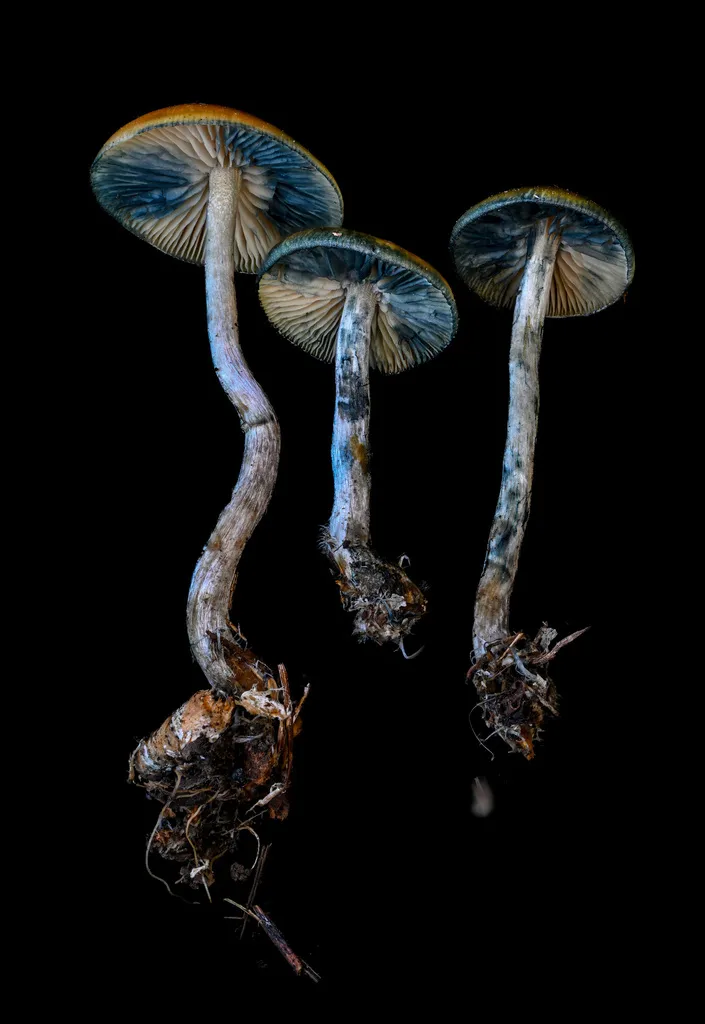
Foto yang diasosiasikan dengan Psilocybe subaeruginosa pada iNaturalist. Identifikasi mengikuti sumber; bukan penentuan spesimen independen.
Kredit dan penggunaan
(c) Alan Rockefeller, some rights reserved (CC BY), uploaded by Alan Rockefeller
Ukuran dan format gambar disesuaikan tanpa pemotongan, retus, atau penambahan skala. Lisensi sumber dipertahankan.
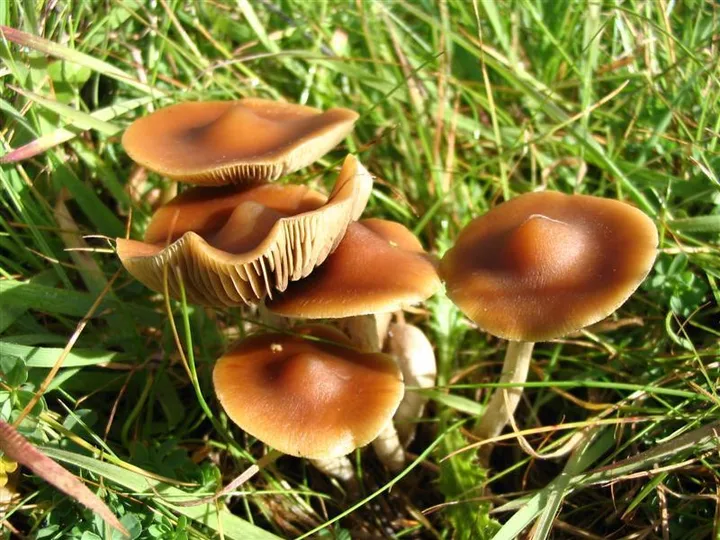
Foto yang diasosiasikan dengan Psilocybe subaeruginosa pada iNaturalist. Identifikasi mengikuti sumber; bukan penentuan spesimen independen.
Kredit dan penggunaan
(c) Alan Rockefeller, some rights reserved (CC BY), uploaded by Alan Rockefeller
Ukuran dan format gambar disesuaikan tanpa pemotongan, retus, atau penambahan skala. Lisensi sumber dipertahankan.
Sumber profil
- [1] Psilocybe subaeruginosa — Wikipedia (en)
- [2] Catalogue of Life Base2026-09-11 — Psilocybe subaeruginosa
- [3] Dokumentasi visual — Psilocybe subaeruginosa
- [4] Dokumentasi visual — Psilocybe subaeruginosa
- [5] Dokumentasi visual — Psilocybe subaeruginosa
Rujukan halaman ini
Fungi. Psilocybe subaeruginosa. Rilis data fungi-2026-10-12-expansion-2. https://fungi-atlas.mgadib.chatgpt.site/jamur/psilocybe-subaeruginosa/.
Status: impor sumber; pemeriksaan editorial per profil belum selesai. Untuk klaim biologis, gunakan rujukan pada bagian terkait dan periksa konteks sumber aslinya.