01 / Morfologi

Ciri dan struktur

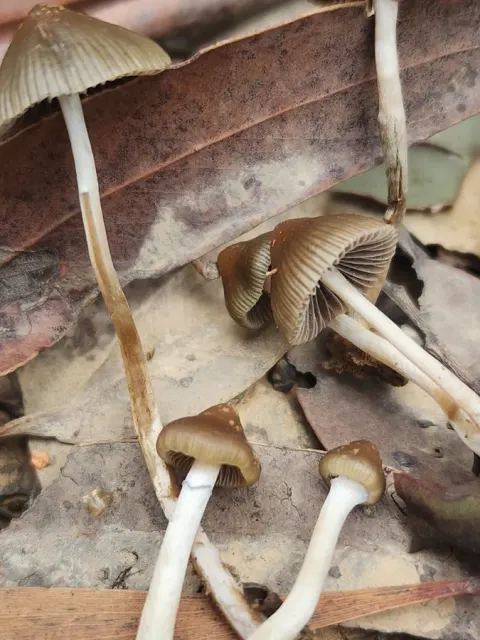
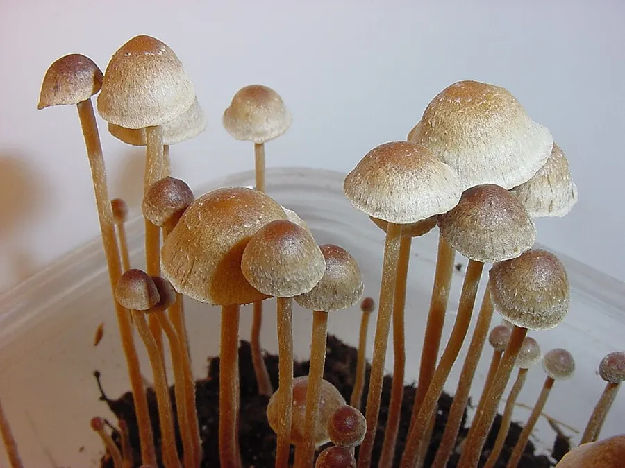
The cap ranges in shape from convex or conic with a slight umbo, expanding in age to become flattened or with a slight central depression; it reaches diameters of 1 – 2.4 cm. The surface is smooth, not striate (grooved), ochraceous brown to straw brown, buff to yellowish-grey when dry, with slight bluish tones at the margin, hygrophanous, and somewhat sticky when wet. The gills are more or less adnate (broadly attached to the stem slightly above the bottom of the gill, with most of the gill fused to the stem) and brown to dark purple brown in color with lighter edges. The stem is 2 – 6 cm long, 1 – 2 mm thick, and equal in width throughout to slightly enlarged near the base. There are fibrils near the top of the stem. The partial veil is cortinate (cobweb-like, similar to the partial veil of Cortinarius species), and soon disappears. The flesh is whitish to yellowish, and bruises blue when injured. The taste and odor are slightly farinaceous (similar to freshly ground flour). The spore print is purple-brown. When viewed with a microscope, the spores of P. tampanensis are somewhat rhombic in face view and roughly elliptical in side view; they have dimensions of 8.8–9.9 by 7–8.8 by 5.5–6.6 μm. Spores appear brownish-yellow when mounted in a solution of potassium hydroxide, and have a thick, smooth wall, a distinct germ pore, and a short appendage. The basidia (spore-bearing cells) are four-spored, hyaline (translucent), and measure 14–22 by 8–10 μm. The cheilocystidia (cystidia on the gill face) measure 16–22 by 4–9 μm, and are lageniform (flask-shaped) with flexuous thin necks that are 2.2–3 μm thick, and infrequently have irregular branches.

Sebagian uraian ditampilkan dalam bahasa sumber; terjemahannya belum tersedia atau belum lolos pemeriksaan.

Rujukan: [1]

Pengukuran mengikuti objek, spesimen atau kultur, dan metode yang disebutkan sumber. Baca uraian asli untuk konteks lengkap.

02 / Kehidupan

Habitat dan peran ekologis

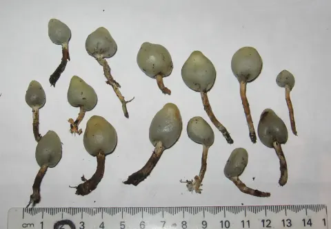
For almost two decades after its discovery, Psilocybe tampanensis was known only from the type locality, southeast of Brandon, Florida. In 1996, Guzmán reported finding it in a meadow with sandy soil in a deciduous forest in Pearl River County, Mississippi, a habitat similar to that of the type location. Like some spesies rumput yang psikoaktif lainnya seperti Psilocybe semilanceata, Conocybe cyanopus, P. tampanensis dapat membentuk sclerotia ⁇ a massa mycelia yang keras yang lebih tahan terhadap kondisi lingkungan yang buruk daripada mycelia normal. Bentuk seperti tartufi ini memberikan jamur perlindungan dari kebakaran hutan dan bencana alam lainnya. Psilocybe species yang dikenal menghasilkan sklerotia lainnya termasuk Psilocybe mexicana dan Psilocybe caerulescens. Sclerotia are juga diproduksi ketika spesies ditanam di budidaya.

Sebagian uraian ditampilkan dalam bahasa sumber; terjemahannya belum tersedia atau belum lolos pemeriksaan.

Rujukan: [1]

Habitat atau konteks temuan
hutan; padang rumput; kultur yang didokumentasikan
Substrat
Informasi belum tersedia pada sumber rilis ini.
Peran atau konteks
Belum dikategorikan pada rilis ini.
Temukan hubungan serupa
03 / Siklus hidup

Reproduksi

The spore print is purple-brown. When viewed with a microscope, the spores of P. tampanensis are somewhat rhombic in face view and roughly elliptical in side view; they have dimensions of 8.8–9.9 by 7–8.8 by 5.5–6.6 μm. Spores appear brownish-yellow when mounted in a solution of potassium hydroxide, and have a thick, smooth wall, a distinct germ pore, and a short appendage. The basidia (spore-bearing cells) are four-spored, hyaline (translucent), and measure 14–22 by 8–10 μm. The cheilocystidia (cystidia on the gill face) measure 16–22 by 4–9 μm, and are lageniform (flask-shaped) with flexuous thin necks that are 2.2–3 μm thick, and infrequently have irregular branches. There are no pleurocystidia (cystidia on the gill face).

Sebagian uraian ditampilkan dalam bahasa sumber; terjemahannya belum tersedia atau belum lolos pemeriksaan.

Rujukan: [1]

04 / Batas informasi

Catatan identitas dan cakupan

  • Nama diterima mengikuti Catalogue of Life Base2026-09-11; uraian mempertahankan konsep dan nama sumber aslinya.
  • Original geographic, specimen and life-cycle scope retained; source import awaits individual editorial review.
  • Impor dan terjemahan otomatis belum diperiksa editorial per profil. Struktur reproduktif yang disebutkan tidak membuktikan seluruh siklus hidup diketahui.
  • Pengukuran pada uraian mengikuti spesimen, kultur, dan metode sumber; angka tidak diperluas ke semua populasi.

Catatan distribusi dalam narasi mengikuti sumbernya. Dokumentasi spesimen atau foto mengikuti catatan sumber. Bukti wilayah Indonesia dan penilaian konservasi tidak disimpulkan dari gambar.

05 / Dokumentasi

Melihat dengan konteks

Dokumentasi Psilocybe tampanensis dari Wikimedia Commons. Psilocybe tampanensis Identifikasi mengikuti record berkas sumber.

Jenis
Fotografi
Kredit
Workman
Sumber media
Buka entri sumber

Gambar menunjukkan objek menurut identifikasi sumber, bukan verifikasi independen spesimen oleh Fungi. Tidak ada skala baru yang ditambahkan.

06 / Rujukan

Sumber profil

  1. [1] Psilocybe tampanensis — Wikipedia (en)Bagian sumber: Frozen revision 1371341593
    Tanggal penghimpunan sumber: 2026-10-12
    Teks sumber: CC BY-SA4.0 · Wikipedia contributors
  2. [2] Catalogue of Life Base2026-09-11 — Psilocybe tampanensisBagian sumber: ColDP NameUsage.tsv ID 4PHDZ; accepted species; source 2073
    Tanggal penghimpunan sumber: 2026-10-12
  3. [3] Dokumentasi visual — Psilocybe tampanensisBagian sumber: Individual media record; identification follows the source, not independent determination
    Tanggal penghimpunan sumber: 2026-10-12
    Media sumber: CC BY-SA 3.0 · Workman

Adaptasi teks: CC BY-SA 4.0. Wikipedia contributors — Psilocybe tampanensis — Wikipedia (en)

Uraian asli dan riwayat sumber

Riwayat atau halaman sumber

Ringkasan sumber

The cap ranges in shape from convex or conic with a slight umbo, expanding in age to become flattened or with a slight central depression; it reaches diameters of 1 – 2.4 cm. The surface is smooth, not striate (grooved), ochraceous brown to straw brown, buff to yellowish-grey when dry, with slight bluish tones at the margin, hygrophanous, and somewhat sticky when wet. The gills are more or less adnate (broadly attached to the stem slightly above the bottom of the gill, with most of the gill fused to the stem) and brown to dark purple brown in color with lighter edges.

Revisi sumber

Morfologi sumber

The cap ranges in shape from convex or conic with a slight umbo, expanding in age to become flattened or with a slight central depression; it reaches diameters of 1 – 2.4 cm. The surface is smooth, not striate (grooved), ochraceous brown to straw brown, buff to yellowish-grey when dry, with slight bluish tones at the margin, hygrophanous, and somewhat sticky when wet. The gills are more or less adnate (broadly attached to the stem slightly above the bottom of the gill, with most of the gill fused to the stem) and brown to dark purple brown in color with lighter edges. The stem is 2 – 6 cm long, 1 – 2 mm thick, and equal in width throughout to slightly enlarged near the base. There are fibrils near the top of the stem. The partial veil is cortinate (cobweb-like, similar to the partial veil of Cortinarius species), and soon disappears. The flesh is whitish to yellowish, and bruises blue when injured. The taste and odor are slightly farinaceous (similar to freshly ground flour). The spore print is purple-brown. When viewed with a microscope, the spores of P. tampanensis are somewhat rhombic in face view and roughly elliptical in side view; they have dimensions of 8.8–9.9 by 7–8.8 by 5.5–6.6 μm. Spores appear brownish-yellow when mounted in a solution of potassium hydroxide, and have a thick, smooth wall, a distinct germ pore, and a short appendage. The basidia (spore-bearing cells) are four-spored, hyaline (translucent), and measure 14–22 by 8–10 μm. The cheilocystidia (cystidia on the gill face) measure 16–22 by 4–9 μm, and are lageniform (flask-shaped) with flexuous thin necks that are 2.2–3 μm thick, and infrequently have irregular branches.

Revisi sumber

Ekologi sumber

For almost two decades after its discovery, Psilocybe tampanensis was known only from the type locality, southeast of Brandon, Florida. In 1996, Guzmán reported finding it in a meadow with sandy soil in a deciduous forest in Pearl River County, Mississippi, a habitat similar to that of the type location. Like some other psychoactive grassland species such as Psilocybe semilanceata, Conocybe cyanopus, P. tampanensis can form sclerotia–a hardened mass of mycelia that is more resistant to adverse environmental conditions than normal mycelia. This truffle-like form gives the fungus some protection from wildfires and other natural disasters. Other Psilocybe species known to produce sclerotia include Psilocybe mexicana and Psilocybe caerulescens. Sclerotia are also produced when the species is grown in culture.

Revisi sumber

Struktur reproduksi sumber

The spore print is purple-brown. When viewed with a microscope, the spores of P. tampanensis are somewhat rhombic in face view and roughly elliptical in side view; they have dimensions of 8.8–9.9 by 7–8.8 by 5.5–6.6 μm. Spores appear brownish-yellow when mounted in a solution of potassium hydroxide, and have a thick, smooth wall, a distinct germ pore, and a short appendage. The basidia (spore-bearing cells) are four-spored, hyaline (translucent), and measure 14–22 by 8–10 μm. The cheilocystidia (cystidia on the gill face) measure 16–22 by 4–9 μm, and are lageniform (flask-shaped) with flexuous thin necks that are 2.2–3 μm thick, and infrequently have irregular branches. There are no pleurocystidia (cystidia on the gill face).

Revisi sumber

Rujukan halaman ini

Fungi. Psilocybe tampanensis. Rilis data fungi-2026-10-12-expansion-2. https://fungi-atlas.mgadib.chatgpt.site/jamur/psilocybe-tampanensis/.

Status: impor sumber; pemeriksaan editorial per profil belum selesai. Untuk klaim biologis, gunakan rujukan pada bagian terkait dan periksa konteks sumber aslinya.