Ciri dan struktur
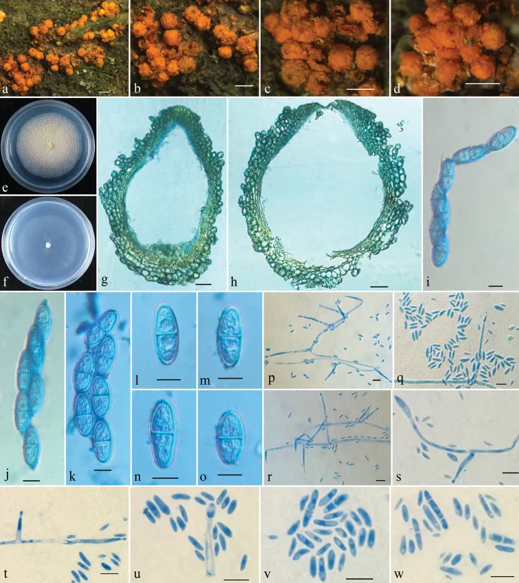
Mycelium not visible around ascomata or on natural substrata. Ascomata superficial, gregarious, with basal stroma, pyriform to subglobose, non-papillate, yellow to orange, often with a darker red ostiolar area when dry, turning dark red in KOH, becoming slightly yellow in LA, 421–549 × 333–470 μm (n = 8). Perithecial surface warted, 30–93 µm thick, of textura globulosa to textura angularis, cells 10–27 × 8–18 µm, walls 1.5–2.5 µm thick. Perithecial wall of two layers, 45–70 µm thick, outer layer 25–45 µm thick, of textura globulosa to textura angularis; inner layer 7–25 µm thick, of textura prismatica. Asci unitunicate, clavate, 8-spored, 93–130 × (11–)15–25 µm (112.6 ± 12.6 × 18.9 ± 3.2 μm). Ascospores ellipsoid to broadly ellipsoid, 1-septate, striate, uniseriate or biseriate above and uniseriate below, hyaline, 20–28 × 8–12 µm (24.0 ± 2.0 × 10.1 ± 0.9 μm). Colony on PDA 42 mm diameter after 7 d under daylight at 25 °C, surface velvety, with white aerial mycelium, producing pale pinkish pigment in medium. Colony on SNA reaches 40 mm diameter after 7 d under daylight at 25 °C, surface with sparse whitish aerial mycelium. Conidiophores simply branched, 18–50 × 2–3 μm. Microconidia allantoid to rod shaped, slightly curved, 0(1–2)-septate, 3–14(–18) × 1.2–2.5(–3) μm (6.7 ± 3.1 × 1.6 ± 0.4 μm).
Habitat dan peran ekologis
Mycelium not visible around ascomata or on natural substrata. Ascomata superficial, gregarious, with basal stroma, pyriform to subglobose, non-papillate, yellow to orange, often with a darker red ostiolar area when dry, turning dark red in KOH, becoming slightly yellow in LA, 421–549 × 333–470 μm (n = 8). Perithecial surface warted, 30–93 µm thick, of textura globulosa to textura angularis, cells 10–27 × 8–18 µm, walls 1.5–2.5 µm thick. Perithecial wall of two layers, 45–70 µm thick, outer layer 25–45 µm thick, of textura globulosa to textura angularis; inner layer 7–25 µm thick, of textura prismatica. Asci unitunicate, clavate, 8-spored, 93–130 × (11–)15–25 µm (112.6 ± 12.6 × 18.9 ± 3.2 μm). Ascospores ellipsoid to broadly ellipsoid, 1-septate, striate, uniseriate or biseriate above and uniseriate below, hyaline, 20–28 × 8–12 µm (24.0 ± 2.0 × 10.1 ± 0.9 μm). Colony on PDA 42 mm diameter after 7 d under daylight at 25 °C, surface velvety, with white aerial mycelium, producing pale pinkish pigment in medium. Colony on SNA reaches 40 mm diameter after 7 d under daylight at 25 °C, surface with sparse whitish aerial mycelium. Conidiophores simply branched, 18–50 × 2–3 μm. Microconidia allantoid to rod shaped, slightly curved, 0(1–2)-septate, 3–14(–18) × 1.2–2.5(–3) μm (6.7 ± 3.1 × 1.6 ± 0.4 μm).
- Habitat atau konteks temuan
- kultur yang didokumentasikan
- Substrat
- Informasi belum tersedia pada sumber rilis ini.
- Peran atau konteks
- Belum dikategorikan pada rilis ini.
Reproduksi
Mycelium not visible around ascomata or on natural substrata. Ascomata superficial, gregarious, with basal stroma, pyriform to subglobose, non-papillate, yellow to orange, often with a darker red ostiolar area when dry, turning dark red in KOH, becoming slightly yellow in LA, 421–549 × 333–470 μm (n = 8). Perithecial surface warted, 30–93 µm thick, of textura globulosa to textura angularis, cells 10–27 × 8–18 µm, walls 1.5–2.5 µm thick. Perithecial wall of two layers, 45–70 µm thick, outer layer 25–45 µm thick, of textura globulosa to textura angularis; inner layer 7–25 µm thick, of textura prismatica. Asci unitunicate, clavate, 8-spored, 93–130 × (11–)15–25 µm (112.6 ± 12.6 × 18.9 ± 3.2 μm).
Catatan identitas dan cakupan
- Nama diterima mengikuti Catalogue of Life Base2026-09-11; uraian mempertahankan konsep dan nama sumber aslinya.
- Preserve original treatment localities, hosts and substrate scope. Materials examined describe documented specimens, not the full global range.
- Impor dan terjemahan otomatis belum diperiksa editorial per profil. Struktur reproduktif yang disebutkan tidak membuktikan seluruh siklus hidup diketahui.
- Pengukuran pada uraian mengikuti spesimen, kultur, dan metode sumber; angka tidak diperluas ke semua populasi.
Catatan distribusi dalam narasi mengikuti sumbernya. Dokumentasi spesimen atau foto mengikuti catatan sumber. Bukti wilayah Indonesia dan penilaian konservasi tidak disimpulkan dari gambar.
Melihat dengan konteks
Rugonectria microconidia a–d ascomata on natural substratum e colony on PDA f colony on SNA g, h median section through perithecium i–k asci with ascospores l–o ascospores p–s conidiophores and conidia t, u conidiogenous cells and conidia v, w microconidia. Scale bars: 0.5 mm (a–d); 50 μm (g, h); 10 μm (i–w).
- Jenis
- Pelat ilmiah
- Kredit
- Zhao-Qing Zeng, Wen-Ying Zhuang
- Lisensi
- CC BY 4.0
- Sumber media
- Buka entri sumber
Sumber profil
- [1] The genera Rugonectria and Thelonectria (Hypocreales, Nectriaceae) in China — Rugonectria microconidia
- [2] Catalogue of Life Base2026-09-11 — Rugonectria microconidia
- [3] Dokumentasi visual — Rugonectria microconidia
Rujukan halaman ini
Fungi. Rugonectria microconidia. Rilis data fungi-2026-10-12-expansion-2. https://fungi-atlas.mgadib.chatgpt.site/jamur/rugonectria-microconidia/.
Status: impor sumber; pemeriksaan editorial per profil belum selesai. Untuk klaim biologis, gunakan rujukan pada bagian terkait dan periksa konteks sumber aslinya.