01 / Morfologi

Ciri dan struktur

Asexual morph: Synnemata arising from the host, 3.5–5 mm long, irregularly branched, conidia in abundance at the apex. Colonies fast-growing on PDA, 35–40 mm in 14 days at 25 °C, floccose, crater-shaped, white to pale pink, sporulating abundantly at the centrum, forming a white concentric ring. Reverse pale brown. Hyphae smooth-walled, branched, septate, hyaline, 1.3–3.1 µm wide. Conidiophores cylindrical, solitary or verticillate, 6.4–10.5 × 1.7–2.1 µm. Phialides verticillate, in whorls of two to five, sometimes solitary on hyphae, 5.6–9.3 µm long, basal portion cylindrical to narrowly lageniform, tapering gradually or abruptly toward the apex, from 1.5–2.1 µm wide (base) to 0.6–1.0 µm wide (apex). Conidia smooth and hyaline, spherical, elliptical or fusiform, one-celled, 2.0–3.1 × 1.3–1.9 µm, often in chains. Size and shape of phialides and conidia similar in culture and on natural substratum. Notes: Regarding phylogenetic relationships, S. sinensis formed a distinct lineage and was closely related to S. hymenopterorum. Morphologically, synnemata were observed in S. sinensis, and synnemata were not observed in S. hymenopterorum. The phialides of S. sinensis (5.6–9.3 µm) were shorter than those of S. hymenopterorum (6.5–10.6 µm). The conidia of S. hymenopterorum were fusiform to ovoid, 1.9–2.5 × 1.5–2.1 µm, but those of S. sinensis were spherical, elliptical or fusiform, 2.0–3.1 × 1.3–1.9 µm. Samsoniella sinensis was also easily distinguished from S. hymenopterorum by its host. Samsoniella sinensis was found to occur on Lepidoptera and Dermaptera, while S. hymenopterorum was only found to occur on Hymenopterous insects.

Sebagian uraian ditampilkan dalam bahasa sumber; terjemahannya belum tersedia atau belum lolos pemeriksaan.

Rujukan: [1]

Pengukuran mengikuti objek, spesimen atau kultur, dan metode yang disebutkan sumber. Baca uraian asli untuk konteks lengkap.

02 / Kehidupan

Habitat dan peran ekologis

Distribution: Currently only known in Kunming City and Chuxiong City, Yunnan Province, China, and Guiyang City, Guizhou Province, China.

Sebagian uraian ditampilkan dalam bahasa sumber; terjemahannya belum tersedia atau belum lolos pemeriksaan.

Rujukan: [1]

Habitat atau konteks temuan
Informasi belum tersedia pada sumber rilis ini.
Substrat
Informasi belum tersedia pada sumber rilis ini.
Peran atau konteks
Belum dikategorikan pada rilis ini.
Temukan hubungan serupa
03 / Siklus hidup

Reproduksi

Asexual morph: Synnemata arising from the host, 3.5–5 mm long, irregularly branched, conidia in abundance at the apex. Colonies fast-growing on PDA, 35–40 mm in 14 days at 25 °C, floccose, crater-shaped, white to pale pink, sporulating abundantly at the centrum, forming a white concentric ring. Reverse pale brown. Hyphae smooth-walled, branched, septate, hyaline, 1.3–3.1 µm wide. Conidiophores cylindrical, solitary or verticillate, 6.4–10.5 × 1.7–2.1 µm. Phialides verticillate, in whorls of two to five, sometimes solitary on hyphae, 5.6–9.3 µm long, basal portion cylindrical to narrowly lageniform, tapering gradually or abruptly toward the apex, from 1.5–2.1 µm wide (base) to 0.6–1.0 µm wide (apex).

Sebagian uraian ditampilkan dalam bahasa sumber; terjemahannya belum tersedia atau belum lolos pemeriksaan.

Rujukan: [1]

04 / Batas informasi

Catatan identitas dan cakupan

  • Nama diterima mengikuti Catalogue of Life Base2026-09-11; uraian mempertahankan konsep dan nama sumber aslinya.
  • Preserve original treatment localities, hosts and substrate scope. Materials examined describe documented specimens, not the full global range.
  • Impor dan terjemahan otomatis belum diperiksa editorial per profil. Struktur reproduktif yang disebutkan tidak membuktikan seluruh siklus hidup diketahui.
  • Pengukuran pada uraian mengikuti spesimen, kultur, dan metode sumber; angka tidak diperluas ke semua populasi.

Catatan distribusi dalam narasi mengikuti sumbernya. Dokumentasi spesimen atau foto mengikuti catatan sumber. Bukti wilayah Indonesia dan penilaian konservasi tidak disimpulkan dari gambar.

05 / Dokumentasi

Melihat dengan konteks

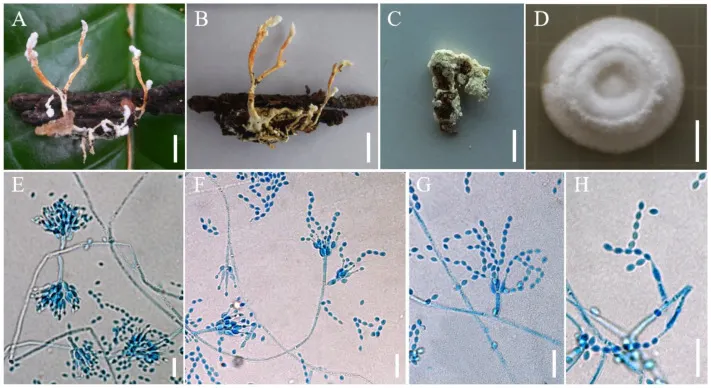
Morphology of Samsoniella sinensis. (A, B) Larva of Lepidoptera infected by S. sinensis. (C) Dermaptera infected by S. sinensis. (D) Culture character on PDA medium. (E–H) Conidiogenous cells (conidiophores, phialides) and conidia on PDA. Scale A, B: 5 mm; C: 3 mm; D: 10 mm; E – H: 10 µm.

Jenis
Pelat ilmiah
Kredit
© 2022 by the authors.
Lisensi
CC BY 4.0
Sumber media
Buka entri sumber

Gambar menunjukkan objek menurut identifikasi sumber, bukan verifikasi independen spesimen oleh Fungi. Tidak ada skala baru yang ditambahkan.

06 / Rujukan

Sumber profil

  1. [1] Morphological and Phylogenetic Characterization Reveals Five New Species of Samsoniella (Cordycipitaceae, Hypocreales) — Samsoniella sinensisBagian sumber: sec3dot2-jof-08-00747_paragraph_46
    Tanggal penghimpunan sumber: 2026-10-12
    Teks sumber: CC BY 4.0 · Wang Z, Wang Y, Dong Q, Fan Q, Dao VM, Yu H.
  2. [2] Catalogue of Life Base2026-09-11 — Samsoniella sinensisBagian sumber: ColDP NameUsage.tsv ID B2QPM; accepted species; source 2073
    Tanggal penghimpunan sumber: 2026-10-12
  3. [3] Dokumentasi visual — Samsoniella sinensisBagian sumber: Individual media record; identification follows the source, not independent determination
    Tanggal penghimpunan sumber: 2026-10-12
    Media sumber: CC BY 4.0 · © 2022 by the authors.

Adaptasi teks: CC BY 4.0. Wang Z, Wang Y, Dong Q, Fan Q, Dao VM, Yu H. — Morphological and Phylogenetic Characterization Reveals Five New Species of Samsoniella (Cordycipitaceae, Hypocreales) — Samsoniella sinensis

Uraian asli dan riwayat sumber

Riwayat atau halaman sumber

Ringkasan sumber

Asexual morph: Synnemata arising from the host, 3.5–5 mm long, irregularly branched, conidia in abundance at the apex. Colonies fast-growing on PDA, 35–40 mm in 14 days at 25 °C, floccose, crater-shaped, white to pale pink, sporulating abundantly at the centrum, forming a white concentric ring. Reverse pale brown. Hyphae smooth-walled, branched, septate, hyaline, 1.3–3.1 µm wide. Conidiophores cylindrical, solitary or verticillate, 6.4–10.5 × 1.7–2.1 µm. Phialides verticillate, in whorls of two to five, sometimes solitary on hyphae, 5.6–9.3 µm long, basal portion cylindrical to narrowly lageniform, tapering gradually or abruptly toward the apex, from 1.5–2.1 µm wide (base) to 0.6–1.0 µm wide (apex).

Revisi sumber

Morfologi sumber

Asexual morph: Synnemata arising from the host, 3.5–5 mm long, irregularly branched, conidia in abundance at the apex. Colonies fast-growing on PDA, 35–40 mm in 14 days at 25 °C, floccose, crater-shaped, white to pale pink, sporulating abundantly at the centrum, forming a white concentric ring. Reverse pale brown. Hyphae smooth-walled, branched, septate, hyaline, 1.3–3.1 µm wide. Conidiophores cylindrical, solitary or verticillate, 6.4–10.5 × 1.7–2.1 µm. Phialides verticillate, in whorls of two to five, sometimes solitary on hyphae, 5.6–9.3 µm long, basal portion cylindrical to narrowly lageniform, tapering gradually or abruptly toward the apex, from 1.5–2.1 µm wide (base) to 0.6–1.0 µm wide (apex). Conidia smooth and hyaline, spherical, elliptical or fusiform, one-celled, 2.0–3.1 × 1.3–1.9 µm, often in chains. Size and shape of phialides and conidia similar in culture and on natural substratum. Notes: Regarding phylogenetic relationships, S. sinensis formed a distinct lineage and was closely related to S. hymenopterorum. Morphologically, synnemata were observed in S. sinensis, and synnemata were not observed in S. hymenopterorum. The phialides of S. sinensis (5.6–9.3 µm) were shorter than those of S. hymenopterorum (6.5–10.6 µm). The conidia of S. hymenopterorum were fusiform to ovoid, 1.9–2.5 × 1.5–2.1 µm, but those of S. sinensis were spherical, elliptical or fusiform, 2.0–3.1 × 1.3–1.9 µm. Samsoniella sinensis was also easily distinguished from S. hymenopterorum by its host. Samsoniella sinensis was found to occur on Lepidoptera and Dermaptera, while S. hymenopterorum was only found to occur on Hymenopterous insects.

Revisi sumber

Ekologi sumber

Distribution: Currently only known in Kunming City and Chuxiong City, Yunnan Province, China, and Guiyang City, Guizhou Province, China.

Revisi sumber

Struktur reproduksi sumber

Asexual morph: Synnemata arising from the host, 3.5–5 mm long, irregularly branched, conidia in abundance at the apex. Colonies fast-growing on PDA, 35–40 mm in 14 days at 25 °C, floccose, crater-shaped, white to pale pink, sporulating abundantly at the centrum, forming a white concentric ring. Reverse pale brown. Hyphae smooth-walled, branched, septate, hyaline, 1.3–3.1 µm wide. Conidiophores cylindrical, solitary or verticillate, 6.4–10.5 × 1.7–2.1 µm. Phialides verticillate, in whorls of two to five, sometimes solitary on hyphae, 5.6–9.3 µm long, basal portion cylindrical to narrowly lageniform, tapering gradually or abruptly toward the apex, from 1.5–2.1 µm wide (base) to 0.6–1.0 µm wide (apex).

Revisi sumber

Rujukan halaman ini

Fungi. Samsoniella sinensis. Rilis data fungi-2026-10-12-expansion-2. https://fungi-atlas.mgadib.chatgpt.site/jamur/samsoniella-sinensis/.

Status: impor sumber; pemeriksaan editorial per profil belum selesai. Untuk klaim biologis, gunakan rujukan pada bagian terkait dan periksa konteks sumber aslinya.