01 / Morfologi

Ciri dan struktur

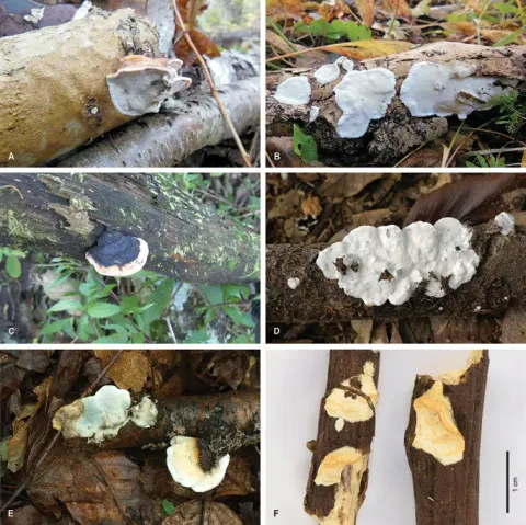
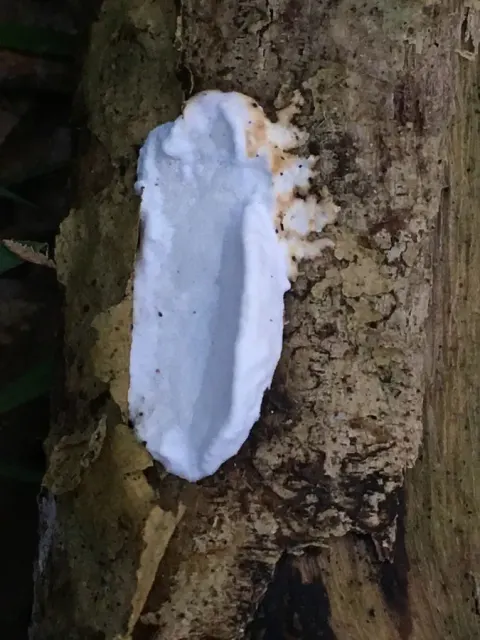
Basidiocarps annual; half-resupinate; pilei up to 3 cm wide, nodulous or up to 4 mm thick and fleshy, blunt edged; upper surface slightly rough, matted, white to cream coloured when young, turning ochraceous; context faintly zonate in longitudinal section with thin dark lines separating layers of growth; tube layer up to 1 mm thick; pores (7–)8–9(–10) per mm. Hyphal structure: skeletal hyphae in context / subiculum (1.0–)2.0–3.3(–4.0) µm wide, in trama (1.0–)2.0–3.5(–4.9) µm wide, generative hyphae in trama 1.0–2.2(–3.0) µm wide. Basidiospores See general description of microscopic structures; (2.8–)2.9–3.0(–3.1)×0.5–0.6 µm, L=2.95 µm, W=0.55 µm, Q’=4.8–6.0, Q=5.36, n=90/3.

Sebagian uraian ditampilkan dalam bahasa sumber; terjemahannya belum tersedia atau belum lolos pemeriksaan.

Rujukan: [1]

Pengukuran mengikuti objek, spesimen atau kultur, dan metode yang disebutkan sumber. Baca uraian asli untuk konteks lengkap.

02 / Kehidupan

Habitat dan peran ekologis

Available material is limited but suggests a temperate East Asian distribution. Russian specimens represent rather small basidiocarps that were collected from thin, fallen angiosperm branches. The holotype from Japan is a sturdier basidiocarp that was probably growing on a thick branch or a log.

Sebagian uraian ditampilkan dalam bahasa sumber; terjemahannya belum tersedia atau belum lolos pemeriksaan.

Rujukan: [1]

Habitat atau konteks temuan
Informasi belum tersedia pada sumber rilis ini.
Substrat
Informasi belum tersedia pada sumber rilis ini.
Peran atau konteks
Belum dikategorikan pada rilis ini.
Temukan hubungan serupa
03 / Siklus hidup

Reproduksi

Basidiocarps annual; half-resupinate; pilei up to 3 cm wide, nodulous or up to 4 mm thick and fleshy, blunt edged; upper surface slightly rough, matted, white to cream coloured when young, turning ochraceous; context faintly zonate in longitudinal section with thin dark lines separating layers of growth; tube layer up to 1 mm thick; pores (7–)8–9(–10) per mm. Hyphal structure: skeletal hyphae in context / subiculum (1.0–)2.0–3.3(–4.0) µm wide, in trama (1.0–)2.0–3.5(–4.9) µm wide, generative hyphae in trama 1.0–2.2(–3.0) µm wide. Basidiospores See general description of microscopic structures; (2.8–)2.9–3.0(–3.1)×0.5–0.6 µm, L=2.95 µm, W=0.55 µm, Q’=4.8–6.0, Q=5.36, n=90/3.

Sebagian uraian ditampilkan dalam bahasa sumber; terjemahannya belum tersedia atau belum lolos pemeriksaan.

Rujukan: [1]

04 / Batas informasi

Catatan identitas dan cakupan

  • Nama diterima mengikuti Catalogue of Life Base2026-09-11; uraian mempertahankan konsep dan nama sumber aslinya.
  • Preserve original treatment localities, hosts and substrate scope. Materials examined describe documented specimens, not the full global range.
  • Impor dan terjemahan otomatis belum diperiksa editorial per profil. Struktur reproduktif yang disebutkan tidak membuktikan seluruh siklus hidup diketahui.
  • Pengukuran pada uraian mengikuti spesimen, kultur, dan metode sumber; angka tidak diperluas ke semua populasi.

Catatan distribusi dalam narasi mengikuti sumbernya. Dokumentasi spesimen atau foto mengikuti catatan sumber. Bukti wilayah Indonesia dan penilaian konservasi tidak disimpulkan dari gambar.

05 / Dokumentasi

Melihat dengan konteks

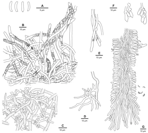
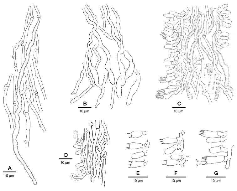
Microscopic structures of the Skeletocutis nivea complex. A S. lepida, tramal skeletal hypha amongst generative hyphae (holotype) B S. semipileata, ends of generative and skeletal hyphae in trama (Miettinen 17135) C S. nemoralis, tube trama and hymenium (holotype) D S. nivea, tube trama and hymenium with encrusted generocystidia (epitype) E S. nivea, basidia (Miettinen 16350) F S. semipileata, basidia (epitype) G S. cummata, the largest basidia in the the S. nivea complex (Niemelä 9088).

Jenis
Pelat ilmiah
Kredit
Aku Korhonen, Jaya Seelan Sathiya Seelan, Otto Miettinen
Lisensi
CC BY 4.0
Sumber media
Buka entri sumber

Gambar menunjukkan objek menurut identifikasi sumber, bukan verifikasi independen spesimen oleh Fungi. Tidak ada skala baru yang ditambahkan.

06 / Rujukan

Sumber profil

  1. [1] Cryptic species diversity in polypores: the Skeletocutis nivea species complex — Skeletocutis lepidaBagian sumber: PMC6060227_section_56
    Tanggal penghimpunan sumber: 2026-10-12
    Teks sumber: CC BY 4.0 · Korhonen, Aku & Seelan, Jaya Seelan Sathiya & Miettinen, Otto
  2. [2] Catalogue of Life Base2026-09-11 — Skeletocutis lepidaBagian sumber: ColDP NameUsage.tsv ID 6Z4GR; accepted species; source 2073
    Tanggal penghimpunan sumber: 2026-10-12
  3. [3] Dokumentasi visual — Skeletocutis lepidaBagian sumber: Individual media record; identification follows the source, not independent determination
    Tanggal penghimpunan sumber: 2026-10-12
    Media sumber: CC BY 4.0 · Aku Korhonen, Jaya Seelan Sathiya Seelan, Otto Miettinen

Adaptasi teks: CC BY 4.0. Korhonen, Aku & Seelan, Jaya Seelan Sathiya & Miettinen, Otto — Cryptic species diversity in polypores: the Skeletocutis nivea species complex — Skeletocutis lepida

Uraian asli dan riwayat sumber

Riwayat atau halaman sumber

Ringkasan sumber

Basidiocarps annual; half-resupinate; pilei up to 3 cm wide, nodulous or up to 4 mm thick and fleshy, blunt edged; upper surface slightly rough, matted, white to cream coloured when young, turning ochraceous; context faintly zonate in longitudinal section with thin dark lines separating layers of growth; tube layer up to 1 mm thick; pores (7–)8–9(–10) per mm. Hyphal structure: skeletal hyphae in context / subiculum (1.0–)2.0–3.3(–4.0) µm wide, in trama (1.0–)2.0–3.5(–4.9) µm wide, generative hyphae in trama 1.0–2.2(–3.0) µm wide.

Revisi sumber

Morfologi sumber

Basidiocarps annual; half-resupinate; pilei up to 3 cm wide, nodulous or up to 4 mm thick and fleshy, blunt edged; upper surface slightly rough, matted, white to cream coloured when young, turning ochraceous; context faintly zonate in longitudinal section with thin dark lines separating layers of growth; tube layer up to 1 mm thick; pores (7–)8–9(–10) per mm. Hyphal structure: skeletal hyphae in context / subiculum (1.0–)2.0–3.3(–4.0) µm wide, in trama (1.0–)2.0–3.5(–4.9) µm wide, generative hyphae in trama 1.0–2.2(–3.0) µm wide. Basidiospores See general description of microscopic structures; (2.8–)2.9–3.0(–3.1)×0.5–0.6 µm, L=2.95 µm, W=0.55 µm, Q’=4.8–6.0, Q=5.36, n=90/3.

Revisi sumber

Ekologi sumber

Available material is limited but suggests a temperate East Asian distribution. Russian specimens represent rather small basidiocarps that were collected from thin, fallen angiosperm branches. The holotype from Japan is a sturdier basidiocarp that was probably growing on a thick branch or a log.

Revisi sumber

Struktur reproduksi sumber

Basidiocarps annual; half-resupinate; pilei up to 3 cm wide, nodulous or up to 4 mm thick and fleshy, blunt edged; upper surface slightly rough, matted, white to cream coloured when young, turning ochraceous; context faintly zonate in longitudinal section with thin dark lines separating layers of growth; tube layer up to 1 mm thick; pores (7–)8–9(–10) per mm. Hyphal structure: skeletal hyphae in context / subiculum (1.0–)2.0–3.3(–4.0) µm wide, in trama (1.0–)2.0–3.5(–4.9) µm wide, generative hyphae in trama 1.0–2.2(–3.0) µm wide. Basidiospores See general description of microscopic structures; (2.8–)2.9–3.0(–3.1)×0.5–0.6 µm, L=2.95 µm, W=0.55 µm, Q’=4.8–6.0, Q=5.36, n=90/3.

Revisi sumber

Rujukan halaman ini

Fungi. Skeletocutis lepida. Rilis data fungi-2026-10-12-expansion-2. https://fungi-atlas.mgadib.chatgpt.site/jamur/skeletocutis-lepida/.

Status: impor sumber; pemeriksaan editorial per profil belum selesai. Untuk klaim biologis, gunakan rujukan pada bagian terkait dan periksa konteks sumber aslinya.