Ciri dan struktur
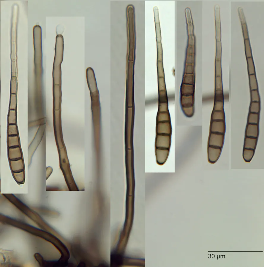
In 1958 I made the new combination Corynespora foveolata because some conidiophores showed percurrent proliferations through the scar of the ter minal conidium with a conidium produced on each successive proliferation. Corynespora is the genus used also by Ellis (1960, 1971) in his redescription of this species. However, close examination shows that the terminal cell of the conidiophore has 5 to 8 scattered pores in the lateral wall. In old herbarium material no conidia could be seen attached at these pores; however, one should expect such multiple lateral production because my preparations. at least, show a far greater number of conidia than of condiophores and their proliferations. Furthermore many conidia have a rounded base rather than a flattened one (with a convex depression) as found on conidia produced terminally. In the New Zealand collections. undisturbed conidiophores had developed within the hollow of the stems and they show several radiating conidia attached to the apical cell of the conidiophore. For this reason I revert to Patouillard's original disposition of this species. Conidiophores in the type collections of Helminthosporium cantonense Sace. and H. microsorum P. Henn. non D. Sacc. also bear pores in the terminal cell of the conidiophores. These names are synonyms of H. foveolatum Matsushima (1975) illustrated Helminthosporium sp. on dead stems of Ph yllostachys edule in Japan with pores in the wall of the upper cell of the conidiophore and conidia with a roughened vall; this is surely H. foveolatum
Habitat dan peran ekologis
On dead stems of Phyllostachys sp. (1. 2) Auckland Prov.. Epsom, 13.1.1963, S.D., POD 20484 (DAOM 93811), DAOM 109319. In 1958 I made the new combination Corynespora foveolata because some conidiophores showed percurrent proliferations through the scar of the ter minal conidium with a conidium produced on each successive proliferation. Corynespora is the genus used also by Ellis (1960, 1971) in his redescription of this species. However, close examination shows that the terminal cell of the conidiophore has 5 to 8 scattered pores in the lateral wall. In old herbarium material no conidia could be seen attached at these pores; however, one should expect such multiple lateral production because my preparations. at least, show a far greater number of conidia than of condiophores and their proliferations. Furthermore many conidia have a rounded base rather than a flattened one (with a convex depression) as found on conidia produced terminally. In the New Zealand collections. undisturbed conidiophores had developed within the hollow of the stems and they show several radiating conidia attached to the apical cell of the conidiophore. For this reason I revert to Patouillard's original disposition of this species.
- Habitat atau konteks temuan
- Informasi belum tersedia pada sumber rilis ini.
- Substrat
- Informasi belum tersedia pada sumber rilis ini.
- Peran atau konteks
- Belum dikategorikan pada rilis ini.
Reproduksi
In 1958 I made the new combination Corynespora foveolata because some conidiophores showed percurrent proliferations through the scar of the ter minal conidium with a conidium produced on each successive proliferation. Corynespora is the genus used also by Ellis (1960, 1971) in his redescription of this species. However, close examination shows that the terminal cell of the conidiophore has 5 to 8 scattered pores in the lateral wall. In old herbarium material no conidia could be seen attached at these pores; however, one should expect such multiple lateral production because my preparations. at least, show a far greater number of conidia than of condiophores and their proliferations. Furthermore many conidia have a rounded base rather than a flattened one (with a convex depression) as found on conidia produced terminally.
Catatan identitas dan cakupan
- Nama diterima mengikuti Catalogue of Life Base2026-09-11; uraian mempertahankan konsep dan nama sumber aslinya.
- Historical description as cited by BiotaNZ. Preserve explicit New Zealand/locality, host and specimen qualifiers. Distribution is the cited source scope, not a current global range.
- Impor dan terjemahan otomatis belum diperiksa editorial per profil. Struktur reproduktif yang disebutkan tidak membuktikan seluruh siklus hidup diketahui.
- Pengukuran pada uraian mengikuti spesimen, kultur, dan metode sumber; angka tidak diperluas ke semua populasi.
Catatan distribusi dalam narasi mengikuti sumbernya. Dokumentasi spesimen atau foto mengikuti catatan sumber. Bukti wilayah Indonesia dan penilaian konservasi tidak disimpulkan dari gambar.
Melihat dengan konteks
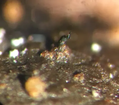
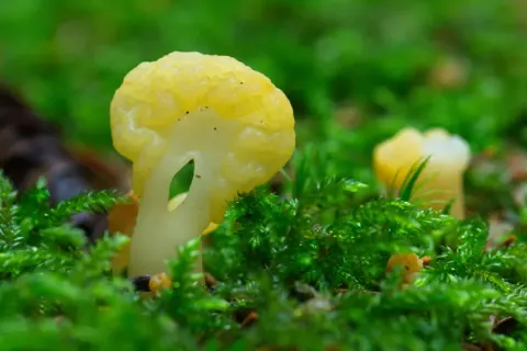
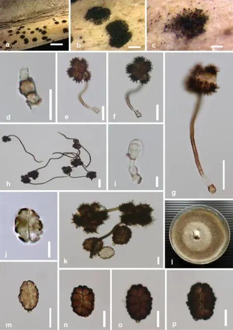
Dokumentasi Solicorynespora foveolata menurut asosiasi foto pada record takson iNaturalist. Kredit: Jerry Cooper. Identitas mengikuti sumber; bukan penentuan spesimen independen.
- Jenis
- Fotografi
- Kredit
- (c) Jerry Cooper, some rights reserved (CC BY), uploaded by Jerry Cooper
- Lisensi
- CC BY 4.0
- Sumber media
- Buka entri sumber
Sumber profil
- [1] Hughes, S.J. 1978: New Zealand fungi. 25. Miscellaneous species. New Zealand Journal of Botany 16(3): 311-370. — Helminthosporium foveolatum
- [2] Catalogue of Life Base2026-09-11 — Solicorynespora foveolata
- [3] Dokumentasi visual — Solicorynespora foveolata
Rujukan halaman ini
Fungi. Solicorynespora foveolata. Rilis data fungi-2026-10-12-expansion-2. https://fungi-atlas.mgadib.chatgpt.site/jamur/solicorynespora-foveolata/.
Status: impor sumber; pemeriksaan editorial per profil belum selesai. Untuk klaim biologis, gunakan rujukan pada bagian terkait dan periksa konteks sumber aslinya.