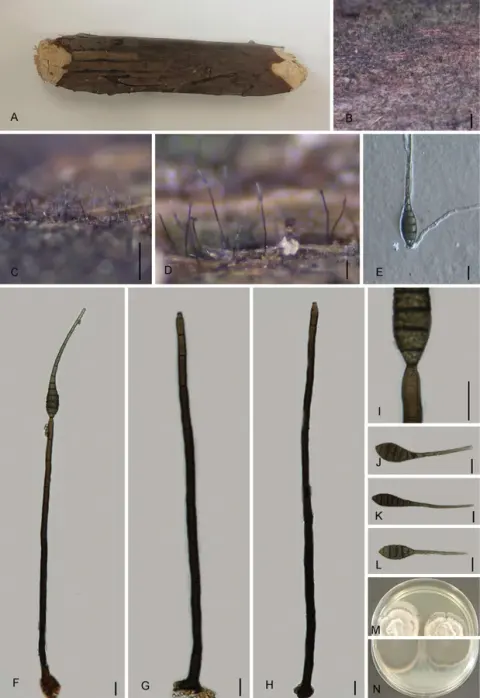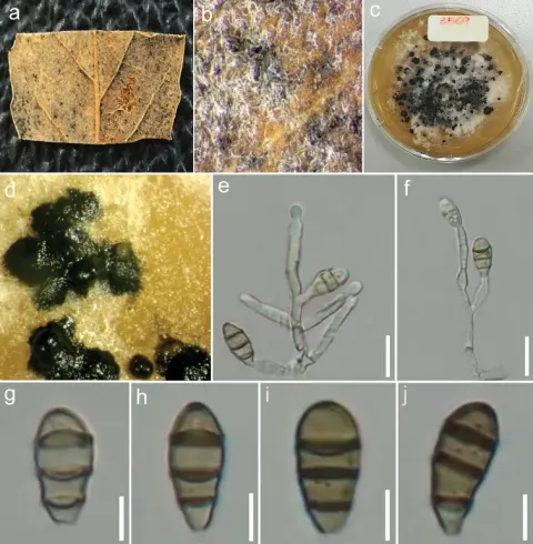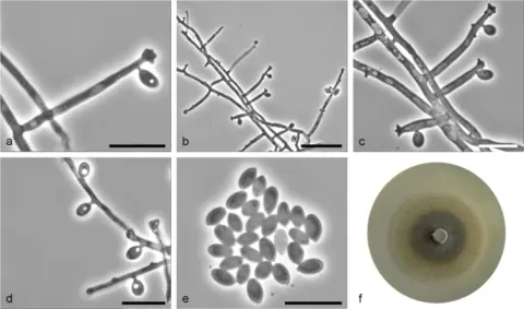
Ciri dan struktur
Sexual morph. Ascomata pseudoperithecial, scattered, immersed, erumpent at the ostiolar neck, subglobose to depressed globose, 480–770 μm high, 530–800 μm diam, ostiolate. Ostiolar neck central, 80–120 μm long, 110–180 μm wide. Ascomatal wall (24–)35–45 μm thick at the sides, composed of 5–6 layers of polygonal, 10–21 × 5–8(–9) μm, brown cells. Pseudoparaphyses 2.5–4 μm wide, septate, branched and anastomosed. Asci bitunicate, clavate, 181–230(–245) × 30–39 μm (mean ± SD = 212.6 ± 13.3 × 34.4 ± 2.2 μm, n = 33), short-stalked (30–70 μm long, mean ± SD = 46.1 ± 11.1 μm, n = 33), 8-spored, rarely 4-, 6- or 7-spored due to meiotic failure or stunted spores, with uni- to bi-seriate ascospores. Ascospores clavate-ellipsoid, 30–45 × 13–18 μm (mean ± SD = 37.0 ± 3.4 × 15.6 ± 1.2 μm, n = 197), l/w = 1.9–3.1(–3.3) (mean ± SD = 2.4 ± 0.3, n = 197), 1-septate, with a submedian septum [(0.55–)0.58–0.67(–0.70), mean ± SD = 0.63 ± 0.03, n = 100], strongly constricted at the septum, brown to dark brown, smooth to faintly roughened, with an entire sheath. Asexual morph. Conidiomata pycnidial, scattered, immersed, ampulliform, 310–560 μm high, 240–450 μm diam, ostiolate. Ostiolar neck central, 80–140 μm long, 100–220 μm wide. Conidiomatal wall 20–47 μm thick, composed of 4–6 layers of polygonal, 7.5–16 × 4.5–8 μm, brown cells. Paraphyses 2.5–3.5 μm wide, septate, branched. Conidiophores absent. Conidiogenous cells annellidic, cylindrical, (12–)15–23(–29) × 3–7.5(–11) μm (mean ± SD = 19.8 ± 5.1 × 5.8 ± 2.0 μm, n = 13). Conidia globose to subglobose, 14.5–22.5 × 14–17 μm (mean ± SD = 18.8 ± 1.7 × 15.1 ± 0.7 μm, n = 102), l/w = 1.1–1.4(–1.6) (mean ± SD = 1.2 ± 0.1, n = 102), aseptate, hyaline, smooth, with thick spore wall (1–1.5 μm).
Habitat dan peran ekologis
Asia [Georgia (Nakhutsrishvili 1986), the Russian Far East (Vasilyeva 1998)]; Europe [Austria (this study), Belgium, Czech Republic, Denmark (Munk 1957), Finland, France, Germany, Hungary, Italy, Lithuania (Treigienė 1999), Luxembourg, Netherlands, Norway (Nordén et al. 2018), Poland (Mułenko et al. 2008), Portugal, Russia (Mel’nik 2011), Spain, Sweden (Eriksson 2014; this study), Switzerland (Tonolo 1956), UK (Pirozynski and Morgan-Jones 1968; Barr 1982; this study), Ukraine]; North America [Canada, USA (Barr 1982)]. Records of countries without references are based on specimens recorded in Cybertruffle (http://www.cybertruffle.org.uk/eng/), GBIF Secretariat (2023), or MyCoPortal (https://www.mycoportal.org/portal/index.php); true species identities of collections outside of Europe require additional studies.
- Habitat atau konteks temuan
- Informasi belum tersedia pada sumber rilis ini.
- Substrat
- Informasi belum tersedia pada sumber rilis ini.
- Peran atau konteks
- Belum dikategorikan pada rilis ini.
Reproduksi
Sexual morph. Ascomata pseudoperithecial, scattered, immersed, erumpent at the ostiolar neck, subglobose to depressed globose, 480–770 μm high, 530–800 μm diam, ostiolate. Ostiolar neck central, 80–120 μm long, 110–180 μm wide. Ascomatal wall (24–)35–45 μm thick at the sides, composed of 5–6 layers of polygonal, 10–21 × 5–8(–9) μm, brown cells. Pseudoparaphyses 2.5–4 μm wide, septate, branched and anastomosed. Asci bitunicate, clavate, 181–230(–245) × 30–39 μm (mean ± SD = 212.6 ± 13.3 × 34.4 ± 2.2 μm, n = 33), short-stalked (30–70 μm long, mean ± SD = 46.1 ± 11.1 μm, n = 33), 8-spored, rarely 4-, 6- or 7-spored due to meiotic failure or stunted spores, with uni- to bi-seriate ascospores. Ascospores clavate-ellipsoid, 30–45 × 13–18 μm (mean ± SD = 37.0 ± 3.4 × 15.6 ± 1.2 μm, n = 197), l/w = 1.9–3.1(–3.3) (mean ± SD = 2.4 ± 0.3, n = 197), 1-septate, with a submedian septum [(0.55–)0.58–0.67(–0.70), mean ± SD = 0.63 ± 0.03, n = 100], strongly constricted at the septum, brown to dark brown, smooth to faintly roughened, with an entire sheath.
Catatan identitas dan cakupan
- Nama diterima mengikuti Catalogue of Life Base2026-09-11; uraian mempertahankan konsep dan nama sumber aslinya.
- Preserve original treatment localities, hosts and substrate scope. Materials examined describe documented specimens, not the full global range.
- Impor dan terjemahan otomatis belum diperiksa editorial per profil. Struktur reproduktif yang disebutkan tidak membuktikan seluruh siklus hidup diketahui.
- Pengukuran pada uraian mengikuti spesimen, kultur, dan metode sumber; angka tidak diperluas ke semua populasi.
Catatan distribusi dalam narasi mengikuti sumbernya. Dokumentasi spesimen atau foto mengikuti catatan sumber. Bukti wilayah Indonesia dan penilaian konservasi tidak disimpulkan dari gambar.
Melihat dengan konteks
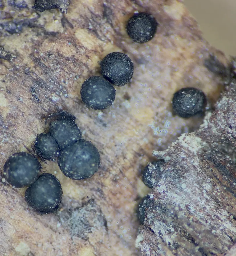
Dokumentasi Splanchnonema ampullaceum menurut asosiasi foto pada record takson iNaturalist. Kredit: Sigrid Jakob. Identitas mengikuti sumber; bukan penentuan spesimen independen.
- Jenis
- Fotografi
- Kredit
- no rights reserved, uploaded by Sigrid Jakob
- Lisensi
- CC0 1.0
- Sumber media
- Buka entri sumber
Dokumentasi tambahan
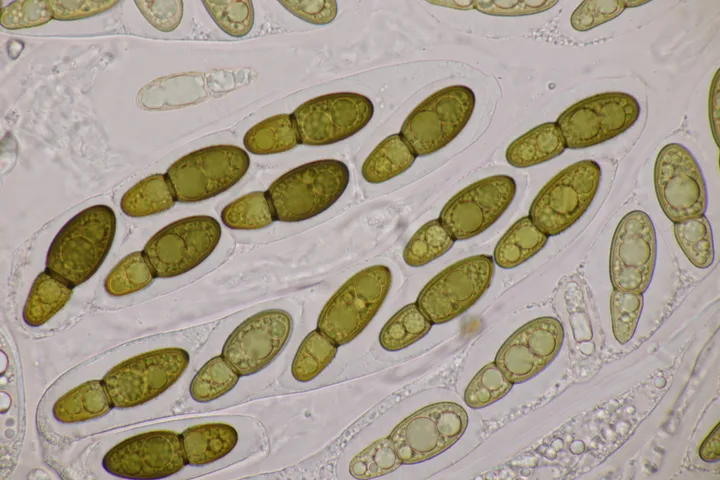
Foto yang diasosiasikan dengan Splanchospora ampullacea pada iNaturalist. Identifikasi mengikuti sumber; bukan penentuan spesimen independen.
Kredit dan penggunaan
(c) Björn Sothmann, some rights reserved (CC BY-SA), uploaded by Björn Sothmann
Ukuran dan format gambar disesuaikan tanpa pemotongan, retus, atau penambahan skala. Lisensi sumber dipertahankan.
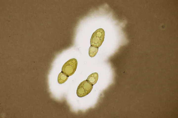
Foto yang diasosiasikan dengan Splanchospora ampullacea pada iNaturalist. Identifikasi mengikuti sumber; bukan penentuan spesimen independen.
Kredit dan penggunaan
(c) Björn Sothmann, some rights reserved (CC BY-SA), uploaded by Björn Sothmann
Ukuran dan format gambar disesuaikan tanpa pemotongan, retus, atau penambahan skala. Lisensi sumber dipertahankan.
Sumber profil
- [1] Revision of the genus Splanchospora (Pleosporales, Neohendersoniaceae) — Splanchospora ampullacea
- [2] Catalogue of Life Base2026-09-11 — Splanchospora ampullacea
- [3] Dokumentasi visual — Splanchospora ampullacea
- [4] Dokumentasi visual — Splanchospora ampullacea
- [5] Dokumentasi visual — Splanchospora ampullacea
Rujukan halaman ini
Fungi. Splanchospora ampullacea. Rilis data fungi-2026-10-12-expansion-2. https://fungi-atlas.mgadib.chatgpt.site/jamur/splanchospora-ampullacea/.
Status: impor sumber; pemeriksaan editorial per profil belum selesai. Untuk klaim biologis, gunakan rujukan pada bagian terkait dan periksa konteks sumber aslinya.