Ciri dan struktur
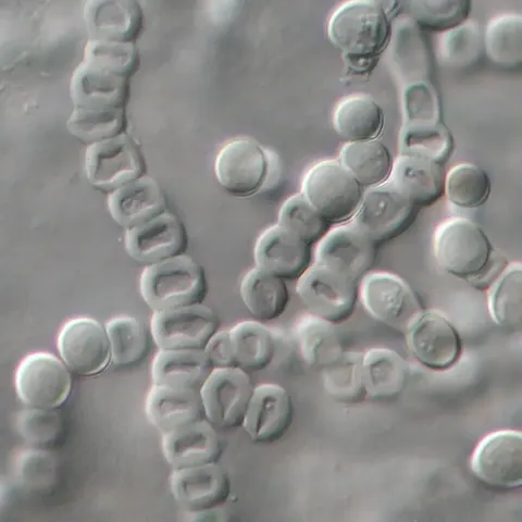
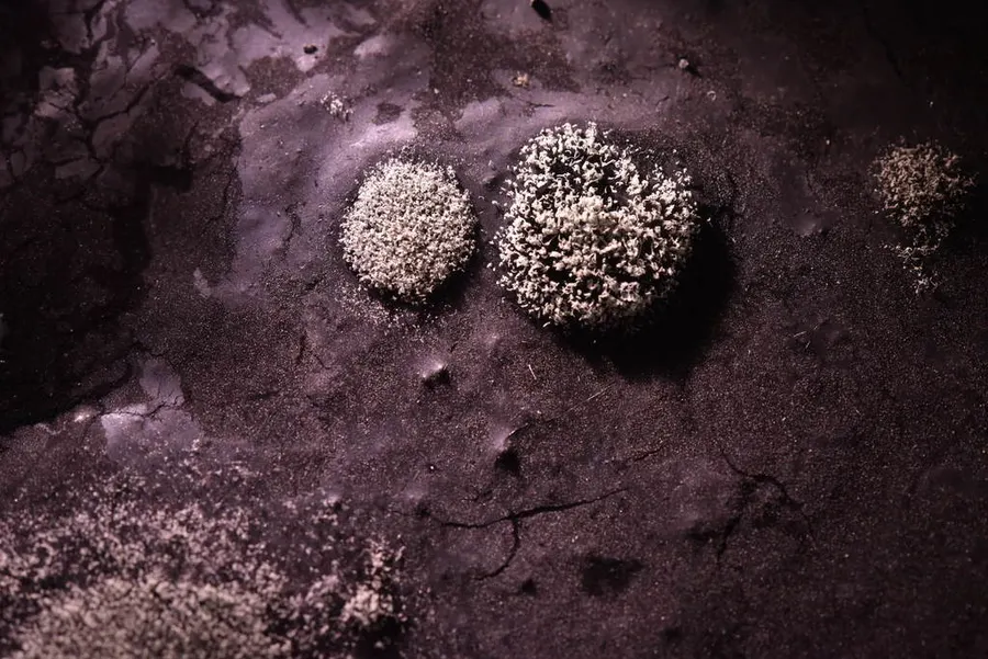
Sporobolomyces salmonicolor produces visible, liposoluble carotenoid pigments, resulting in salmon-pink colonies. The colony surface is smooth and has a pasty texture. There is considerable cell and colony morphology when S. salmonicolor is grown in culture. The budding yeast-like cells produced during the asexual stage are ellipsoidal to subcylindrical and 8–25 × 2–5.5 μm. They can occur singly or in pairs. The ballistoconidia are kidney-shaped and can range in size from 6–18 × 2.5–7.0 μm. The characteristic ballistoconidia are borne by extension of the sterigmata which can reach up to 50 μm in length. Both pseudohyphae and true hyphae may also be present. In its sexual state, Sporobolomyces salmonicolor produces dikaryotic hyphae with clamp connections. At the terminal end of the hyphae, thick-walled teliospores are produced. Teliospores are 9–15 μm in diameter, brown, spherical, and contain lipid-rich globules. Upon germination of the teliospore, basidia with basidiospores are produced. Basidia are transversely septate, two-celled and 4–6 x 20–25 μm in size. Each basidium will generally produce two large basidiospores that are 5–6 × 7–10 μm in size. Prior to the production of the basidium, endospores have also been known to form in the interior of teliospores. This phenomenon is associated with the production of meiospores within the teliospore cytoplasm, ultimately released by rupture of the teliospore wall.
Habitat dan peran ekologis
Sporobolomyces salmonicolor has a broad geographical distribution. It has been isolated from many areas across the world including Europe, North and South America, Asia, Africa and Antarctica where it is known from a broad spectrum of substrates. It is principally characterized as a phyllosphere fungus and is commonly found in areas of decaying organic material such as leaves and grains as well as ripening grapes. Isolates of this species however have been recovered from freshwater, marine water and clinical specimens. It has also been isolated from agricultural areas and indoor built environments. In agricultural environments, Sporobolomyces salmonicolor can pose a respiratory hazard for agricultural workers. Agricultural workers are subject to increased exposure when they partake in activities involving the handling of grains. Sporobolomyces salmonicolor has additionally been isolated from straw in hay barns or hay lofts. Workers in these settings should consider the proper use of masks to avoid infection. If individuals show atopic symptoms, a change in occupation might be considered. In indoor environments, S. salmonicolor has been associated with severe water and mould damage.
- Habitat atau konteks temuan
- Informasi belum tersedia pada sumber rilis ini.
- Substrat
- Informasi belum tersedia pada sumber rilis ini.
- Peran atau konteks
- Belum dikategorikan pada rilis ini.
Reproduksi
Sporobolomyces salmonicolor produces visible, liposoluble carotenoid pigments, resulting in salmon-pink colonies. The colony surface is smooth and has a pasty texture. There is considerable cell and colony morphology when S. salmonicolor is grown in culture. The budding yeast-like cells produced during the asexual stage are ellipsoidal to subcylindrical and 8–25 × 2–5.5 μm. They can occur singly or in pairs. The ballistoconidia are kidney-shaped and can range in size from 6–18 × 2.5–7.0 μm. The characteristic ballistoconidia are borne by extension of the sterigmata which can reach up to 50 μm in length. Both pseudohyphae and true hyphae may also be present. In its sexual state, Sporobolomyces salmonicolor produces dikaryotic hyphae with clamp connections.
Catatan identitas dan cakupan
- Nama diterima mengikuti Catalogue of Life Base2026-09-11; uraian mempertahankan konsep dan nama sumber aslinya.
- Original geographic, specimen and life-cycle scope retained; source import awaits individual editorial review.
- Impor dan terjemahan otomatis belum diperiksa editorial per profil. Struktur reproduktif yang disebutkan tidak membuktikan seluruh siklus hidup diketahui.
- Pengukuran pada uraian mengikuti spesimen, kultur, dan metode sumber; angka tidak diperluas ke semua populasi.
Catatan distribusi dalam narasi mengikuti sumbernya. Dokumentasi spesimen atau foto mengikuti catatan sumber. Bukti wilayah Indonesia dan penilaian konservasi tidak disimpulkan dari gambar.
Melihat dengan konteks
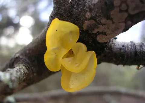
Dokumentasi Sporobolomyces salmonicolor menurut asosiasi foto pada record takson iNaturalist. Kredit: Alan Rockefeller. Identitas mengikuti sumber; bukan penentuan spesimen independen.
- Jenis
- Fotografi
- Kredit
- (c) Alan Rockefeller, some rights reserved (CC BY), uploaded by Alan Rockefeller
- Lisensi
- CC BY 4.0
- Sumber media
- Buka entri sumber
Sumber profil
- [1] Sporobolomyces salmonicolor — Wikipedia (en)
- [2] Catalogue of Life Base2026-09-11 — Sporobolomyces salmonicolor
- [3] Dokumentasi visual — Sporobolomyces salmonicolor
Rujukan halaman ini
Fungi. Sporobolomyces salmonicolor. Rilis data fungi-2026-10-12-expansion-2. https://fungi-atlas.mgadib.chatgpt.site/jamur/sporobolomyces-salmonicolor/.
Status: impor sumber; pemeriksaan editorial per profil belum selesai. Untuk klaim biologis, gunakan rujukan pada bagian terkait dan periksa konteks sumber aslinya.