Ciri dan struktur
Saprobic on leaves of Anthoxanthum odoratum L., visible as black spots, occurring on the upper surface of entire leaf. Sexual morph. Ascomata 110–150 × 90–140 µm (x– = 140–125 µm, n = 10), scattered, solitary, immersed, uniloculate, globose, black. Ostiole 35–40 µm (x– = 39 µm, n = 10) wide, papillate, central, periphysate. Periphyses 15–20 µm long, hyaline. Peridium comprising 2–4 layers of brown to dark brown, thick-walled, cells of textura angularis to textura globularis. Hamathecium comprising 2–4 µm wide, cellular, hyaline, branched, septate, pseudoparaphyses, constricted at the septa, anastomosing mostly above the asci and embedded in a mucilaginous matrix. Asci 85–125 × 20–35 µm (x– = 100 × 30 µm, n = 20), 8-spored, few, bitunicate, fissitunicate, subglobose to clavate, short pedicellate, apically rounded, with an ocular chamber, arising from the base of the ascoma and attached to parenchymatous cell matrix at base. Ascospores 30–35 × 6–9 µm (x– = 35 × 7 µm, n = 25), bi-seriate to tri-seriate, narrowly fusiform, narrowing towards the end cells, reddish to dark brown, 6-septate, second septum supra-median, slightly constricted, not constricted at other septa, second segment swollen, straight, with 12–16 longitudinal furrows on surface, lacking a mucilaginous sheath. Asexual morph. Undetermined. 2 cm diameter after 4 weeks incubated in dark at 25 °C on MEA, pinkish-white, circular, slightly woolly, margin lobate, effuse, lacking aerial mycelium, tightly attached to the media.
Habitat dan peran ekologis
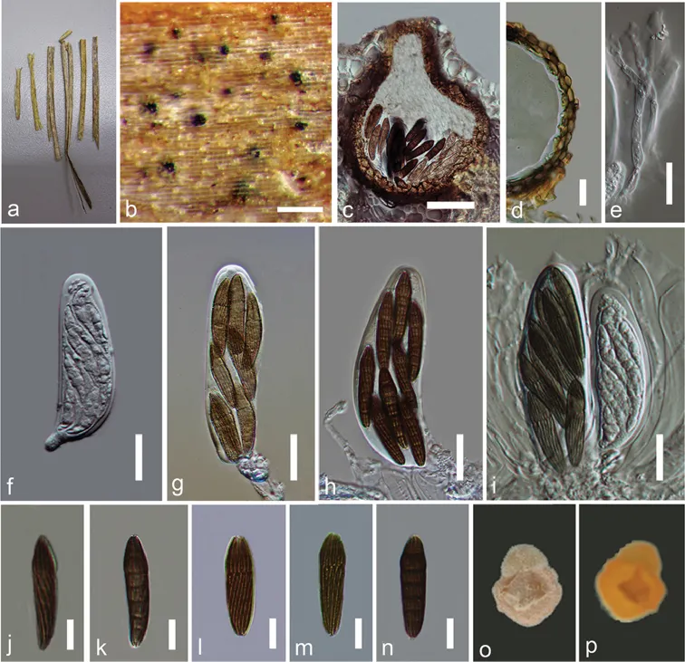
ITALY. Province of Forli-Cesena, Premilcuore, Passodella Valbura, on dead leaves of Anthoxanthum odoratum L. (Poaceae), 25 May 2013, Erio Camporesi, IT 1306 (MFLU 15–0038, holotype; HKAS 83865, paratype): living cultures, MFLUCC 14–0995. Saprobic on leaves of Anthoxanthum odoratum L., visible as black spots, occurring on the upper surface of entire leaf. Sexual morph. Ascomata 110–150 × 90–140 µm (x– = 140–125 µm, n = 10), scattered, solitary, immersed, uniloculate, globose, black. Ostiole 35–40 µm (x– = 39 µm, n = 10) wide, papillate, central, periphysate. Periphyses 15–20 µm long, hyaline. Peridium comprising 2–4 layers of brown to dark brown, thick-walled, cells of textura angularis to textura globularis. Hamathecium comprising 2–4 µm wide, cellular, hyaline, branched, septate, pseudoparaphyses, constricted at the septa, anastomosing mostly above the asci and embedded in a mucilaginous matrix. Asci 85–125 × 20–35 µm (x– = 100 × 30 µm, n = 20), 8-spored, few, bitunicate, fissitunicate, subglobose to clavate, short pedicellate, apically rounded, with an ocular chamber, arising from the base of the ascoma and attached to parenchymatous cell matrix at base. Ascospores 30–35 × 6–9 µm (x– = 35 × 7 µm, n = 25), bi-seriate to tri-seriate, narrowly fusiform, narrowing towards the end cells, reddish to dark brown, 6-septate, second septum supra-median, slightly constricted, not constricted at other septa, second segment swollen, straight, with 12–16 longitudinal furrows on surface, lacking a mucilaginous sheath.
- Habitat atau konteks temuan
- Informasi belum tersedia pada sumber rilis ini.
- Substrat
- Informasi belum tersedia pada sumber rilis ini.
- Peran atau konteks
- Pengurai
Reproduksi
Saprobic on leaves of Anthoxanthum odoratum L., visible as black spots, occurring on the upper surface of entire leaf. Sexual morph. Ascomata 110–150 × 90–140 µm (x– = 140–125 µm, n = 10), scattered, solitary, immersed, uniloculate, globose, black. Ostiole 35–40 µm (x– = 39 µm, n = 10) wide, papillate, central, periphysate. Periphyses 15–20 µm long, hyaline. Peridium comprising 2–4 layers of brown to dark brown, thick-walled, cells of textura angularis to textura globularis. Hamathecium comprising 2–4 µm wide, cellular, hyaline, branched, septate, pseudoparaphyses, constricted at the septa, anastomosing mostly above the asci and embedded in a mucilaginous matrix. Asci 85–125 × 20–35 µm (x– = 100 × 30 µm, n = 20), 8-spored, few, bitunicate, fissitunicate, subglobose to clavate, short pedicellate, apically rounded, with an ocular chamber, arising from the base of the ascoma and attached to parenchymatous cell matrix at base.
Catatan identitas dan cakupan
- Nama diterima mengikuti Catalogue of Life Base2026-09-11; uraian mempertahankan konsep dan nama sumber aslinya.
- Preserve original treatment localities, hosts and substrate scope. Materials examined describe documented specimens, not the full global range.
- Impor dan terjemahan otomatis belum diperiksa editorial per profil. Struktur reproduktif yang disebutkan tidak membuktikan seluruh siklus hidup diketahui.
- Pengukuran pada uraian mengikuti spesimen, kultur, dan metode sumber; angka tidak diperluas ke semua populasi.
Catatan distribusi dalam narasi mengikuti sumbernya. Dokumentasi spesimen atau foto mengikuti catatan sumber. Bukti wilayah Indonesia dan penilaian konservasi tidak disimpulkan dari gambar.
Melihat dengan konteks
Sulcispora supratumida (MFLU 15–0038). a Leaves of Anthoxanthum odoratum b Appearance of ascomata on host surface c Cross section of ascoma d Peridium e Pseudoparaphyses f–i Asci j–n Ascospores o Upper surface of the culture p Lower surface of the culture. Scale bars: 200 µm (b), 50 µm (c), 20 µm (d–i), 10 µm (j–n).
- Jenis
- Pelat ilmiah
- Kredit
- Indunil C. Senanayake, Rajesh Jeewon, Erio Camporesi, Kevin D. Hyde, Yu-Jia Zeng, Sheng-Li Tian, Ning Xie
- Lisensi
- CC BY 4.0
- Sumber media
- Buka entri sumber
Sumber profil
- [1] Sulcispora supratumida sp. nov. (Phaeosphaeriaceae, Pleosporales) on Anthoxanthum odoratum from Italy — Sulcispora supratumida
- [2] Catalogue of Life Base2026-09-11 — Sulcispora supratumida
- [3] Dokumentasi visual — Sulcispora supratumida
Rujukan halaman ini
Fungi. Sulcispora supratumida. Rilis data fungi-2026-10-12-expansion-2. https://fungi-atlas.mgadib.chatgpt.site/jamur/sulcispora-supratumida/.
Status: impor sumber; pemeriksaan editorial per profil belum selesai. Untuk klaim biologis, gunakan rujukan pada bagian terkait dan periksa konteks sumber aslinya.