Saprobic on herbaceous litter. Morph seksual tidak ditentukan. Aseksual morph Colonies terlihat sebagai hitam padat, berambut, bubuk pada inang. Mycelium 1.5–3.5 μm lebar, sebagian terendam pada permukaan pada substrat, terdiri dari bersepta, bercabang, cokelat terang hifa. Conidiophores 9–18.5 × 3–5 μm (\(\bar{x} \) = 14 × 4 μm, n = 50), macronematous to semi- macronematous, mononematous, erect, straight, or slightly flexuous, without apical branches, light brown to brown, ellipsoid to subcylindrical, 0–1-septate, smooth-walled, with 1–3, globlose to subglobse cells, arising from prostrate hyphae.
Sebagian uraian ditampilkan dalam bahasa sumber; terjemahannya belum tersedia atau belum lolos pemeriksaan.
Profil ini diimpor dari uraian spesies pada sumber berlisensi dan sebagian teks diterjemahkan otomatis. Pemeriksaan editorial per profil belum selesai. Bagian yang belum diterjemahkan atau belum lolos pemeriksaan terjemahan tetap ditampilkan dalam bahasa sumber. Angka dan nama ilmiah dipertahankan.
Saprobic on herbaceous litter. Sexual morph Undetermined. Asexual morph Colonies visible as black dense, velvety, powdery on host. Mycelium 1.5–3.5 μm in wide, partly immersed to superficial on the substrate, composed of septate, branched, light brown hyphae. Conidiophores 9–18.5 × 3–5 μm (\(\bar{x} \) = 14 × 4 μm, n = 50), macronematous to semi- macronematous, mononematous, erect, straight, or slightly flexuous, without apical branches, light brown to brown, ellipsoid to subcylindrical, 0–1-septate, smooth-walled, with 1–3, globlose to subglobse cells, arising from prostrate hyphae. Conidiogenous cells 5.5–8.5 × 5–8 μm (\(\bar{x} \) = 7 × 6.5 μm, n = 30), mono- to polyblastic, integrated, terminal or intercalary, cupulate to globose or subglobose, brown to dark brown, smooth to verrucose thick-walled. Conidia (34–)40–70(–77) × 6–8(–10) μm (\(\bar{x} \) = 53.9 × 6.9 μm, n = 50), catenated, composed of moniliform cells, acrogenous, phragmosporous to scolecosporous, brown to dark brown, verruculose, wider cells in the middle, often smaller at apex, 4–20-septate, constricted at the septa, chiefly subcylindrical, produced branched chain, globlose to subglobose of each cell, basal cell slightly truncate, with a darkened scar at the base. Conidial secession schizolytic. Culture characteristics: Colones on PDA, reaching around 40 mm diam., after 2 weeks at room temperature (20–25 °C), colonies medium dense, irregular, slightly effuse, rough, with standing tufts at the centre, umbonate edge, colonies from above, white to cream at the margin, pinkish white to white-brown at the centre, gradually turn to brown when mature, from below white to pink-white at the margin, irregular radiating with pink-brown at the middle, reddish brown to pinkish brown at the centre; not producing pigment in PDA.
Sebagian uraian ditampilkan dalam bahasa sumber; terjemahannya belum tersedia atau belum lolos pemeriksaan.
Pengukuran mengikuti objek, spesimen atau kultur, dan metode yang disebutkan sumber. Baca uraian asli untuk konteks lengkap.
02 / Kehidupan
Habitat dan peran ekologis
Saprobic on herbaceous litter. Sexual morph Undetermined. Asexual morph Colonies visible as black dense, velvety, powdery on host. Mycelium 1.5–3.5 μm in wide, partly immersed to superficial on the substrate, composed of septate, branched, light brown hyphae. Conidiophores 9–18.5 × 3–5 μm (\(\bar{x} \) = 14 × 4 μm, n = 50), macronematous to semi- macronematous, mononematous, erect, straight, or slightly flexuous, without apical branches, light brown to brown, ellipsoid to subcylindrical, 0–1-septate, smooth-walled, with 1–3, globlose to subglobse cells, arising from prostrate hyphae. Conidiogenous cells 5.5–8.5 × 5–8 μm (\(\bar{x} \) = 7 × 6.5 μm, n = 30), mono- to polyblastic, integrated, terminal or intercalary, cupulate to globose or subglobose, brown to dark brown, smooth to verrucose thick-walled. Conidia (34–)40–70(–77) × 6–8(–10) μm (\(\bar{x} \) = 53.9 × 6.9 μm, n = 50), catenated, composed of moniliform cells, acrogenous, phragmosporous to scolecosporous, brown to dark brown, verruculose, wider cells in the middle, often smaller at apex, 4–20-septate, constricted at the septa, chiefly subcylindrical, produced branched chain, globlose to subglobose of each cell, basal cell slightly truncate, with a darkened scar at the base.
Sebagian uraian ditampilkan dalam bahasa sumber; terjemahannya belum tersedia atau belum lolos pemeriksaan.
Saprobic on herbaceous litter. Sexual morph Undetermined. Asexual morph Colonies visible as black dense, velvety, powdery on host. Mycelium 1.5–3.5 μm in wide, partly immersed to superficial on the substrate, composed of septate, branched, light brown hyphae. Conidiophores 9–18.5 × 3–5 μm (\(\bar{x} \) = 14 × 4 μm, n = 50), macronematous to semi- macronematous, mononematous, erect, straight, or slightly flexuous, without apical branches, light brown to brown, ellipsoid to subcylindrical, 0–1-septate, smooth-walled, with 1–3, globlose to subglobse cells, arising from prostrate hyphae. Conidiogenous cells 5.5–8.5 × 5–8 μm (\(\bar{x} \) = 7 × 6.5 μm, n = 30), mono- to polyblastic, integrated, terminal or intercalary, cupulate to globose or subglobose, brown to dark brown, smooth to verrucose thick-walled.
Sebagian uraian ditampilkan dalam bahasa sumber; terjemahannya belum tersedia atau belum lolos pemeriksaan.
Nama diterima mengikuti Catalogue of Life Base2026-09-11; uraian mempertahankan konsep dan nama sumber aslinya.
Preserve original monograph treatment localities, hosts and specimen/substrate scope. Materials examined document specimens, not the full global range.
Impor dan terjemahan otomatis belum diperiksa editorial per profil. Struktur reproduktif yang disebutkan tidak membuktikan seluruh siklus hidup diketahui.
Pengukuran pada uraian mengikuti spesimen, kultur, dan metode sumber; angka tidak diperluas ke semua populasi.
Catatan distribusi dalam narasi mengikuti sumbernya. Dokumentasi spesimen atau foto mengikuti catatan sumber. Bukti wilayah Indonesia dan penilaian konservasi tidak disimpulkan dari gambar.
05 / Dokumentasi
Melihat dengan konteks
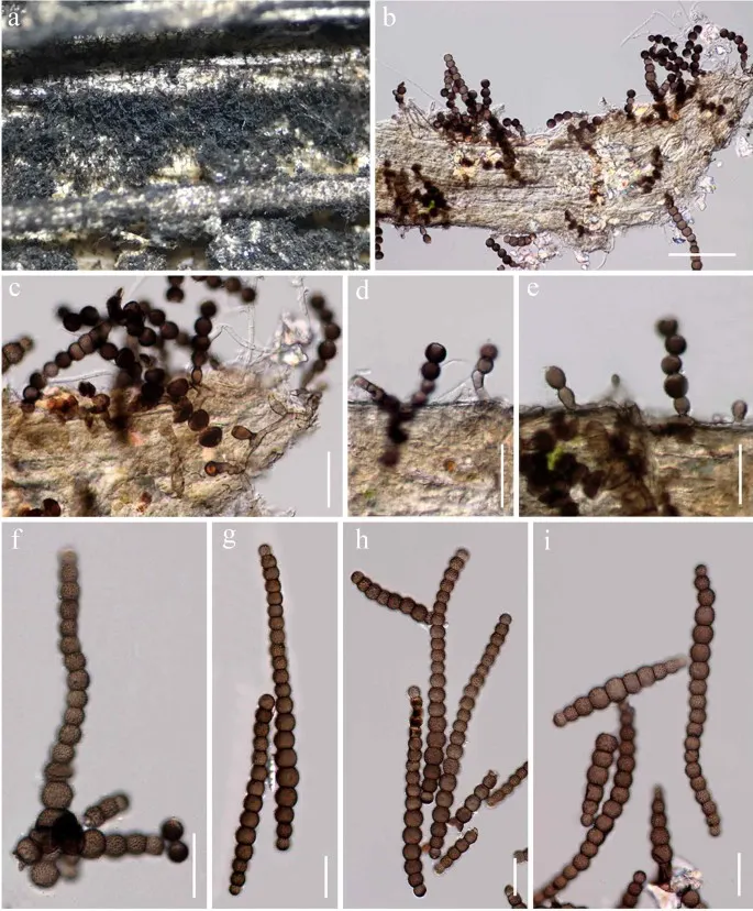
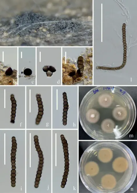
Torula camporesii (MFLU 20-0070, holotype). a Appearance of colonies on host substrate. b, c Colonies on host substrate showing partly immersed mycelium and its erect conidiophores attached with conidia. d, e Conidiophores and conidiogenous cells. f – i Conidia. Scale bars: b = 50 μm, c – i = 20 μm
Jenis
Pelat ilmiah
Kredit
Penulis dan sumber artikel: Hyde, Kevin D.; Dong, Yang; Phookamsak, Rungtiwa; Jeewon, Rajesh; Bhat, D. Jayarama; Jones, E. B. Gareth; Liu, Ning-Guo; Abeywickrama, Pranami D.; Mapook, Ausana; Wei, Deping; Perera, Rekhani H.; Manawasinghe, Ishara S.; Pem, Dhandevi; Bundhun, Digvijayini; Karunarathna, Anuruddha; Ekanayaka, Anusha H.; Bao, Dan-Feng; Li, Junfu; Samarakoon, Milan C.; Chaiwan, Napalai; Lin, Chuan-Gen; Phutthacharoen, Kunthida; Zhang, Sheng-Nan; Senanayake, Indunil C.; Goonasekara, Ishani D.; Thambugala, Kasun M.; Phukhamsakda, Chayanard; Tennakoon, Danushka S.; Jiang, Hong-Bo; Yang, Jing; Zeng, Ming; Huanraluek, Naruemon; Liu, Jian-Kui (Jack); Wijesinghe, Subodini N.; Tian, Qing; Tibpromma, Saowaluck; Brahmanage, Rashika S.; Boonmee, Saranyaphat; Huang, Shi-Ke; Thiyagaraja, Vinodhini; Lu, Yong-Zhong; Jayawardena, Ruvishika S.; Dong, Wei; Yang, Er-Fu; Singh, Sanjay K.; Singh, Shiv Mohan; Rana, Shiwali; Lad, Sneha S.; Anand, Garima; Devadatha, Bandarupalli; Niranjan, M.; Sarma, V. Venkateswara; Liimatainen, Kare; Aguirre-Hudson, Begoña; Niskanen, Tuula; Overall, Andy; Alvarenga, Renato Lúcio Mendes; Gibertoni, Tatiana Baptista; Pfliegler, Walter P.; Horváth, Enikő; Imre, Alexandra; Alves, Amanda Lucia; da Silva Santos, Ana Carla; Tiago, Patricia Vieira; Bulgakov, Timur S.; Wanasinghe, Dhanushaka N.; Bahkali, Ali H.; Doilom, Mingkwan; Elgorban, Abdallah M.; Maharachchikumbura, Sajeewa S. N.; Rajeshkumar, Kunhiraman C.; Haelewaters, Danny; Mortimer, Peter E.; Zhao, Qi; Lumyong, Saisamorn; Xu, Jianchu; Sheng, Jun
Saprobic on herbaceous litter.
Sexual morph Undetermined.
Asexual morph Colonies visible as black dense, velvety, powdery on host.
Mycelium 1.5–3.5 μm in wide, partly immersed to superficial on the substrate, composed of septate, branched, light brown hyphae.
Conidiophores 9–18.5 × 3–5 μm (\(\bar{x} \) = 14 × 4 μm, n = 50), macronematous to semi- macronematous, mononematous, erect, straight, or slightly flexuous, without apical branches, light brown to brown, ellipsoid to subcylindrical, 0–1-septate, smooth-walled, with 1–3, globlose to subglobse cells, arising from prostrate hyphae.
Saprobic on herbaceous litter.
Sexual morph Undetermined.
Asexual morph Colonies visible as black dense, velvety, powdery on host.
Mycelium 1.5–3.5 μm in wide, partly immersed to superficial on the substrate, composed of septate, branched, light brown hyphae.
Conidiophores 9–18.5 × 3–5 μm (\(\bar{x} \) = 14 × 4 μm, n = 50), macronematous to semi- macronematous, mononematous, erect, straight, or slightly flexuous, without apical branches, light brown to brown, ellipsoid to subcylindrical, 0–1-septate, smooth-walled, with 1–3, globlose to subglobse cells, arising from prostrate hyphae.
Conidiogenous cells 5.5–8.5 × 5–8 μm (\(\bar{x} \) = 7 × 6.5 μm, n = 30), mono- to polyblastic, integrated, terminal or intercalary, cupulate to globose or subglobose, brown to dark brown, smooth to verrucose thick-walled.
Conidia (34–)40–70(–77) × 6–8(–10) μm (\(\bar{x} \) = 53.9 × 6.9 μm, n = 50), catenated, composed of moniliform cells, acrogenous, phragmosporous to scolecosporous, brown to dark brown, verruculose, wider cells in the middle, often smaller at apex, 4–20-septate, constricted at the septa, chiefly subcylindrical, produced branched chain, globlose to subglobose of each cell, basal cell slightly truncate, with a darkened scar at the base.
Conidial secession schizolytic.
Culture characteristics: Colones on PDA, reaching around 40 mm diam., after 2 weeks at room temperature (20–25 °C), colonies medium dense, irregular, slightly effuse, rough, with standing tufts at the centre, umbonate edge, colonies from above, white to cream at the margin, pinkish white to white-brown at the centre, gradually turn to brown when mature, from below white to pink-white at the margin, irregular radiating with pink-brown at the middle, reddish brown to pinkish brown at the centre; not producing pigment in PDA.
Saprobic on herbaceous litter.
Sexual morph Undetermined.
Asexual morph Colonies visible as black dense, velvety, powdery on host.
Mycelium 1.5–3.5 μm in wide, partly immersed to superficial on the substrate, composed of septate, branched, light brown hyphae.
Conidiophores 9–18.5 × 3–5 μm (\(\bar{x} \) = 14 × 4 μm, n = 50), macronematous to semi- macronematous, mononematous, erect, straight, or slightly flexuous, without apical branches, light brown to brown, ellipsoid to subcylindrical, 0–1-septate, smooth-walled, with 1–3, globlose to subglobse cells, arising from prostrate hyphae.
Conidiogenous cells 5.5–8.5 × 5–8 μm (\(\bar{x} \) = 7 × 6.5 μm, n = 30), mono- to polyblastic, integrated, terminal or intercalary, cupulate to globose or subglobose, brown to dark brown, smooth to verrucose thick-walled.
Conidia (34–)40–70(–77) × 6–8(–10) μm (\(\bar{x} \) = 53.9 × 6.9 μm, n = 50), catenated, composed of moniliform cells, acrogenous, phragmosporous to scolecosporous, brown to dark brown, verruculose, wider cells in the middle, often smaller at apex, 4–20-septate, constricted at the septa, chiefly subcylindrical, produced branched chain, globlose to subglobose of each cell, basal cell slightly truncate, with a darkened scar at the base.
Saprobic on herbaceous litter.
Sexual morph Undetermined.
Asexual morph Colonies visible as black dense, velvety, powdery on host.
Mycelium 1.5–3.5 μm in wide, partly immersed to superficial on the substrate, composed of septate, branched, light brown hyphae.
Conidiophores 9–18.5 × 3–5 μm (\(\bar{x} \) = 14 × 4 μm, n = 50), macronematous to semi- macronematous, mononematous, erect, straight, or slightly flexuous, without apical branches, light brown to brown, ellipsoid to subcylindrical, 0–1-septate, smooth-walled, with 1–3, globlose to subglobse cells, arising from prostrate hyphae.
Conidiogenous cells 5.5–8.5 × 5–8 μm (\(\bar{x} \) = 7 × 6.5 μm, n = 30), mono- to polyblastic, integrated, terminal or intercalary, cupulate to globose or subglobose, brown to dark brown, smooth to verrucose thick-walled.
Status: impor sumber; pemeriksaan editorial per profil belum selesai. Untuk klaim biologis, gunakan rujukan pada bagian terkait dan periksa konteks sumber aslinya.
Saprobic on tenggelam kayu yang membusuk. Morph seksual: tidak ditentukan. Morph asexual: Koloni menumpahkan pada substrat alami, rapi, berbulu, cokelat. Mycelium immersed ke permukaan, hialin, bersepta, bercabang hifa. Conidiophores…
Saprobic on anggur mati dari Lonicera japonica Thunb (Caprifoliaceae). Morph seksual: tidak ditentukan. Transformasi aseksual: Hipomiketous. Koloni menumpahkan pada substrat alami, tersebar, hitam, dikelompokkan pada substrat. Mycelium…
Saprobic on Malus sp. Morph seksual tidak ditentukan. Aseksual morph Koloni terendam ke permukaan pada substrat inang, cokelat gelap ke hitam, langka, sulit untuk melihat pada inang. Mycelium 0.8–1.5 μm lebar, terdiri dari bercabang,…