Ciri dan struktur
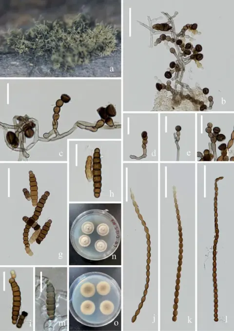
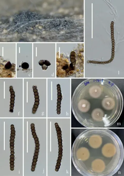
Saprobic on dead Artemisia carvifolia stems. Sexual morph Undetermined. Asexual morph: Colonies effuse on the natural substrate, scattered, hairy, dark brown to black. Mycelium mostly immersed. Conidiophores 16–28 (–45) × 3–4 μm (x̄ = 26 × 4 μm, n = 10), macronematous mononematous, subcylindrical, erect, septate, smooth, straight or slightly flexuous, brown to dark brown, the uppermost side of a transverse compartment is concave inward. Conidiogenous cells 8–10 × 5–7 μm (x̄ = 9 × 6 μm, n =15), holoblastic, polyblastic, doliiform to ellipsoid, dark brown, smooth. Conidia (16–) 25–48 (–70) × 6–10 μm (x̄ = 35 × 8 μm, n = 50), phragmosporous, in branched chains, acrogenous, dry, brown to dark brown, subhyaline at terminal cell, and central cells are significantly larger than both end cells, nearly ellipsoid, constricted at septa, verrucose, easily separating, 2–16-septate, cells subglobose. Conidia germinating on PDA within 12 h. and germ tubes produced at the side. Mycelium superficial, branched, septate, hyaline, smooth. After two weeks of incubation at room temperature, colony edges are irregularly ellipsoid, center is white with gray fuzzy protrusions on the sides, and a translucent gelatinous substance at the outermost periphery. Hyphae flocculent, velvety.
Habitat dan peran ekologis
Saprobic on dead Artemisia carvifolia stems. Sexual morph Undetermined. Asexual morph: Colonies effuse on the natural substrate, scattered, hairy, dark brown to black. Mycelium mostly immersed. Conidiophores 16–28 (–45) × 3–4 μm (x̄ = 26 × 4 μm, n = 10), macronematous mononematous, subcylindrical, erect, septate, smooth, straight or slightly flexuous, brown to dark brown, the uppermost side of a transverse compartment is concave inward. Conidiogenous cells 8–10 × 5–7 μm (x̄ = 9 × 6 μm, n =15), holoblastic, polyblastic, doliiform to ellipsoid, dark brown, smooth. Conidia (16–) 25–48 (–70) × 6–10 μm (x̄ = 35 × 8 μm, n = 50), phragmosporous, in branched chains, acrogenous, dry, brown to dark brown, subhyaline at terminal cell, and central cells are significantly larger than both end cells, nearly ellipsoid, constricted at septa, verrucose, easily separating, 2–16-septate, cells subglobose. Conidia germinating on PDA within 12 h. and germ tubes produced at the side. Mycelium superficial, branched, septate, hyaline, smooth. After two weeks of incubation at room temperature, colony edges are irregularly ellipsoid, center is white with gray fuzzy protrusions on the sides, and a translucent gelatinous substance at the outermost periphery.
- Habitat atau konteks temuan
- kultur yang didokumentasikan
- Substrat
- Informasi belum tersedia pada sumber rilis ini.
- Peran atau konteks
- Pengurai
Reproduksi
Saprobic on dead Artemisia carvifolia stems. Sexual morph Undetermined. Asexual morph: Colonies effuse on the natural substrate, scattered, hairy, dark brown to black. Mycelium mostly immersed. Conidiophores 16–28 (–45) × 3–4 μm (x̄ = 26 × 4 μm, n = 10), macronematous mononematous, subcylindrical, erect, septate, smooth, straight or slightly flexuous, brown to dark brown, the uppermost side of a transverse compartment is concave inward. Conidiogenous cells 8–10 × 5–7 μm (x̄ = 9 × 6 μm, n =15), holoblastic, polyblastic, doliiform to ellipsoid, dark brown, smooth. Conidia (16–) 25–48 (–70) × 6–10 μm (x̄ = 35 × 8 μm, n = 50), phragmosporous, in branched chains, acrogenous, dry, brown to dark brown, subhyaline at terminal cell, and central cells are significantly larger than both end cells, nearly ellipsoid, constricted at septa, verrucose, easily separating, 2–16-septate, cells subglobose.
Catatan identitas dan cakupan
- Nama diterima mengikuti Catalogue of Life Base2026-09-11; uraian mempertahankan konsep dan nama sumber aslinya.
- Preserve original treatment localities, hosts and substrate scope. Materials examined describe documented specimens, not the full global range.
- Impor dan terjemahan otomatis belum diperiksa editorial per profil. Struktur reproduktif yang disebutkan tidak membuktikan seluruh siklus hidup diketahui.
- Pengukuran pada uraian mengikuti spesimen, kultur, dan metode sumber; angka tidak diperluas ke semua populasi.
Catatan distribusi dalam narasi mengikuti sumbernya. Dokumentasi spesimen atau foto mengikuti catatan sumber. Bukti wilayah Indonesia dan penilaian konservasi tidak disimpulkan dari gambar.
Melihat dengan konteks
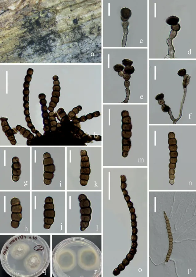
Torula masonii (KUNCC 22-12429) A fungal structure on the substratum C–F conidiophore with conidiogenous cells B, G–O conidia P germinating conidium Q, R colonies on PDA from surface and reverse. Scale bars: 20 μm (B), 10 μm (C–N), 25 μm (O, P)
- Jenis
- Pelat ilmiah
- Kredit
- Wen-Peng Wang, Hong-Wei Shen, Dan-Feng Bao, Yong-Zhong Lu, Qiu-Xia Yang, Xi-Jun Su, Zong-Long Luo
- Lisensi
- CC BY 4.0
- Sumber media
- Buka entri sumber
Dokumentasi tambahan
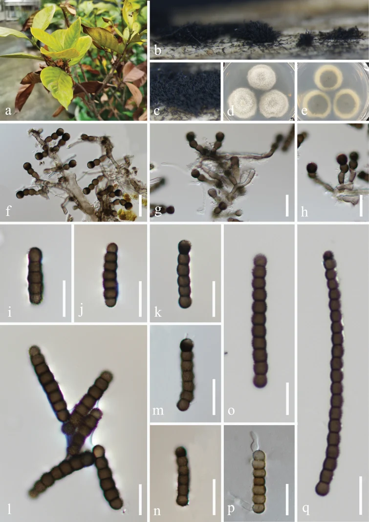
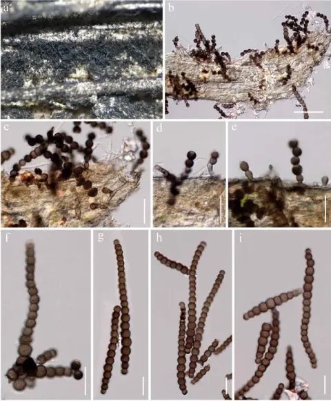
Torula masonii (GZAAS 25-0020, new host record). a. Host Gardenia jasminoides; b, c. Colonies on the host surface; d, e. Colonies on PDA, above (d) and below (e); f−h. Conidiophores and conidiogenous cells; i−o, q. Conidia; p. Germinated conidium. Scale bars: 20 μm (f−h); 15 μm (i−n, p); 10 μm (o, q).
Kredit dan penggunaan
Na Wu, Mei-Feng Chi, Hong-Zhi Du, K. W. Thilini Chethana, Kitiphong Khongphinitbunjong, Ya-Ya Chen, Kevin D. Hyde, Fatimah Al-Otibi, Asha J. Dissanayake, Jian-Kui Liu
Ukuran dan format gambar disesuaikan tanpa pemotongan, retus, atau penambahan skala. Lisensi sumber dipertahankan.
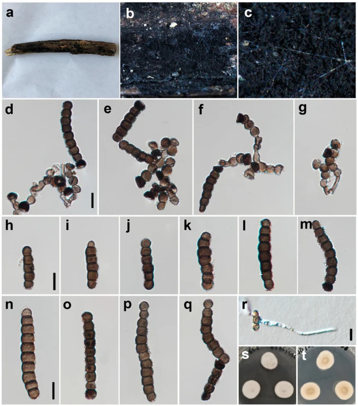
Torula masonii (HUEST 22.0090). (a – c) Colonies on the natural substrate. (d – g) Conidiophores, conidiogenous cells and conidia. (h – q) Conidia; (r) Germinated conidium. (s, t) Culture on PDA from surface and reverse. Scale bars: (d – r) = 10 μm. Scale bar of (d) applies to (d – g). Scale bar of (h) applies to (h – m). Scale bar of (n) applies to (n – q).
Kredit dan penggunaan
© 2023 by the authors.
Ukuran dan format gambar disesuaikan tanpa pemotongan, retus, atau penambahan skala. Lisensi sumber dipertahankan.
Sumber profil
- [1] Two novel species and three new records of Torulaceae from Yunnan Province, China — Torula masonii
- [2] Catalogue of Life Base2026-09-11 — Torula masonii
- [3] Dokumentasi visual — Torula masonii
- [4] Dokumentasi visual — Torula masonii
- [5] Dokumentasi visual — Torula masonii
Rujukan halaman ini
Fungi. Torula masonii. Rilis data fungi-2026-10-12-expansion-2. https://fungi-atlas.mgadib.chatgpt.site/jamur/torula-masonii/.
Status: impor sumber; pemeriksaan editorial per profil belum selesai. Untuk klaim biologis, gunakan rujukan pada bagian terkait dan periksa konteks sumber aslinya.