01 / Morfologi

Ciri dan struktur

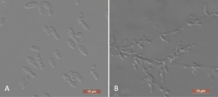
The cells are ovoid to elongate (2–2.5 × 3–8.5 μm) and occur singly or in pairs after being placed in YM broth for three days at 25 °C (Figure 4A). Budding is multilateral. After three days of growth on YM agar at 25 °C, the colonies are white to cream-colored, buttery, and smooth, with entire margins. After two weeks at 25 °C on a Dalmau plate culture with CM agar, pseudohyphae consisting of elongated cells with lateral buds are formed (Figure 4B). True hyphae are not observed. Asci or signs of conjugation are not observed on sporulation media. Fermentation of sugars is absent. Glucose, galactose, l-sorbose, d-glucosamine, d-ribose, d-xylose, l-arabinose, d-arabinose, sucrose, maltose, trehalose, methyl α-d-glucoside, cellobiose, salicin, arbutin, lactose, melezitose, inulin, glycerol, erythritol, ribitol, d-glucitol, d-mannitol, d-gluconate, dl-lactate, succinate, citrate, and ethanol are assimilated. No growth is observed in l-rhamnose, melibiose, raffinose, xylitol, galactitol, myo -inositol, d-glucono-1, 5-lactone, 2-keto-d-gluconate, d-glucuronate, or methanol. In nitrogen-assimilation tests, growth is present on ethylamine, l-lysine, glucosamine, and d-tryptophan, while growth is absent on nitrate, nitrite, cadaverine, creatine, creatinine, and imidazole. Growth is observed at 37 °C but not at 40 °C. Growth in the presence of 0.01% cycloheximide, 10% NaCl with 5% glucose and 1% acetic acid is absent. Starch-like compounds are not produced. Urease activity and diazonium blue B reactions are negative.

Sebagian uraian ditampilkan dalam bahasa sumber; terjemahannya belum tersedia atau belum lolos pemeriksaan.

Rujukan: [1]

Pengukuran mengikuti objek, spesimen atau kultur, dan metode yang disebutkan sumber. Baca uraian asli untuk konteks lengkap.

02 / Kehidupan

Habitat dan peran ekologis

Two strains representing Y. paraaseri were clustered in a well-supported clade and were phylogenetically related to C. aaseri . Yamadazyma paraaseri can be distinguished from C. aaseri based on ITS and D1/D2 LSU loci (8/573 in ITS and 8/531 in D1/D2 LSU). Physiologically, the ability to assimilate d-glucosamine and inulin and the inability to assimilate xylitol and d-glucono-1, 5-lactone are the primary differences between Y. paraaseri and its closest relative, C. aaseri. Additionally, C. aaseri can grow in 10% NaCl with 5% glucose, while Y. paraaseri cannot (Table 2) (Lachance et al. 2011).

Sebagian uraian ditampilkan dalam bahasa sumber; terjemahannya belum tersedia atau belum lolos pemeriksaan.

Rujukan: [1]

Habitat atau konteks temuan
Informasi belum tersedia pada sumber rilis ini.
Substrat
Informasi belum tersedia pada sumber rilis ini.
Peran atau konteks
Belum dikategorikan pada rilis ini.
Temukan hubungan serupa
03 / Siklus hidup

Reproduksi

The cells are ovoid to elongate (2–2.5 × 3–8.5 μm) and occur singly or in pairs after being placed in YM broth for three days at 25 °C (Figure 4A). Budding is multilateral. After three days of growth on YM agar at 25 °C, the colonies are white to cream-colored, buttery, and smooth, with entire margins. After two weeks at 25 °C on a Dalmau plate culture with CM agar, pseudohyphae consisting of elongated cells with lateral buds are formed (Figure 4B). True hyphae are not observed. Asci or signs of conjugation are not observed on sporulation media. Fermentation of sugars is absent. Glucose, galactose, l-sorbose, d-glucosamine, d-ribose, d-xylose, l-arabinose, d-arabinose, sucrose, maltose, trehalose, methyl α-d-glucoside, cellobiose, salicin, arbutin, lactose, melezitose, inulin, glycerol, erythritol, ribitol, d-glucitol, d-mannitol, d-gluconate, dl-lactate, succinate, citrate, and ethanol are assimilated.

Sebagian uraian ditampilkan dalam bahasa sumber; terjemahannya belum tersedia atau belum lolos pemeriksaan.

Rujukan: [1]

04 / Batas informasi

Catatan identitas dan cakupan

  • Nama diterima mengikuti Catalogue of Life Base2026-09-11; uraian mempertahankan konsep dan nama sumber aslinya.
  • Preserve original treatment localities, hosts and substrate scope. Materials examined describe documented specimens, not the full global range.
  • Impor dan terjemahan otomatis belum diperiksa editorial per profil. Struktur reproduktif yang disebutkan tidak membuktikan seluruh siklus hidup diketahui.
  • Pengukuran pada uraian mengikuti spesimen, kultur, dan metode sumber; angka tidak diperluas ke semua populasi.

Catatan distribusi dalam narasi mengikuti sumbernya. Dokumentasi spesimen atau foto mengikuti catatan sumber. Bukti wilayah Indonesia dan penilaian konservasi tidak disimpulkan dari gambar.

05 / Dokumentasi

Melihat dengan konteks

Yamadazyma paraaseri (NYNU 1811114, holotype) A budding cells after three days in YM broth at 25 °C B pseudohyphae on cornmeal agar after two weeks at 25 °C. Scale bars: 10 μm.

Jenis
Pelat ilmiah
Kredit
Wan-Li Gao, Ying Li, Chun-Yue Chai, Zhen-Li Yan, Feng-Li Hui
Lisensi
CC BY 4.0
Sumber media
Buka entri sumber

Gambar menunjukkan objek menurut identifikasi sumber, bukan verifikasi independen spesimen oleh Fungi. Tidak ada skala baru yang ditambahkan.

06 / Rujukan

Sumber profil

  1. [1] New species of Yamadazyma from rotting wood in China — Yamadazyma paraaseriBagian sumber: PMC8413266_section_23
    Tanggal penghimpunan sumber: 2026-10-12
    Teks sumber: CC BY 4.0 · Gao WL, Li Y, Chai CY, Yan ZL, Hui FL.
  2. [2] Catalogue of Life Base2026-09-11 — Yamadazyma paraaseriBagian sumber: ColDP NameUsage.tsv ID B2MCM; accepted species; source 2073
    Tanggal penghimpunan sumber: 2026-10-12
  3. [3] Dokumentasi visual — Yamadazyma paraaseriBagian sumber: Individual media record; identification follows the source, not independent determination
    Tanggal penghimpunan sumber: 2026-10-12
    Media sumber: CC BY 4.0 · Wan-Li Gao, Ying Li, Chun-Yue Chai, Zhen-Li Yan, Feng-Li Hui

Adaptasi teks: CC BY 4.0. Gao WL, Li Y, Chai CY, Yan ZL, Hui FL. — New species of Yamadazyma from rotting wood in China — Yamadazyma paraaseri

Uraian asli dan riwayat sumber

Riwayat atau halaman sumber

Ringkasan sumber

The cells are ovoid to elongate (2–2.5 × 3–8.5 μm) and occur singly or in pairs after being placed in YM broth for three days at 25 °C (Figure 4A). Budding is multilateral. After three days of growth on YM agar at 25 °C, the colonies are white to cream-colored, buttery, and smooth, with entire margins. After two weeks at 25 °C on a Dalmau plate culture with CM agar, pseudohyphae consisting of elongated cells with lateral buds are formed (Figure 4B). True hyphae are not observed. Asci or signs of conjugation are not observed on sporulation media.

Revisi sumber

Morfologi sumber

The cells are ovoid to elongate (2–2.5 × 3–8.5 μm) and occur singly or in pairs after being placed in YM broth for three days at 25 °C (Figure 4A). Budding is multilateral. After three days of growth on YM agar at 25 °C, the colonies are white to cream-colored, buttery, and smooth, with entire margins. After two weeks at 25 °C on a Dalmau plate culture with CM agar, pseudohyphae consisting of elongated cells with lateral buds are formed (Figure 4B). True hyphae are not observed. Asci or signs of conjugation are not observed on sporulation media. Fermentation of sugars is absent. Glucose, galactose, l-sorbose, d-glucosamine, d-ribose, d-xylose, l-arabinose, d-arabinose, sucrose, maltose, trehalose, methyl α-d-glucoside, cellobiose, salicin, arbutin, lactose, melezitose, inulin, glycerol, erythritol, ribitol, d-glucitol, d-mannitol, d-gluconate, dl-lactate, succinate, citrate, and ethanol are assimilated. No growth is observed in l-rhamnose, melibiose, raffinose, xylitol, galactitol, myo -inositol, d-glucono-1, 5-lactone, 2-keto-d-gluconate, d-glucuronate, or methanol. In nitrogen-assimilation tests, growth is present on ethylamine, l-lysine, glucosamine, and d-tryptophan, while growth is absent on nitrate, nitrite, cadaverine, creatine, creatinine, and imidazole. Growth is observed at 37 °C but not at 40 °C. Growth in the presence of 0.01% cycloheximide, 10% NaCl with 5% glucose and 1% acetic acid is absent. Starch-like compounds are not produced. Urease activity and diazonium blue B reactions are negative.

Revisi sumber

Ekologi sumber

Two strains representing Y. paraaseri were clustered in a well-supported clade and were phylogenetically related to C. aaseri . Yamadazyma paraaseri can be distinguished from C. aaseri based on ITS and D1/D2 LSU loci (8/573 in ITS and 8/531 in D1/D2 LSU). Physiologically, the ability to assimilate d-glucosamine and inulin and the inability to assimilate xylitol and d-glucono-1, 5-lactone are the primary differences between Y. paraaseri and its closest relative, C. aaseri. Additionally, C. aaseri can grow in 10% NaCl with 5% glucose, while Y. paraaseri cannot (Table 2) (Lachance et al. 2011).

Revisi sumber

Struktur reproduksi sumber

The cells are ovoid to elongate (2–2.5 × 3–8.5 μm) and occur singly or in pairs after being placed in YM broth for three days at 25 °C (Figure 4A). Budding is multilateral. After three days of growth on YM agar at 25 °C, the colonies are white to cream-colored, buttery, and smooth, with entire margins. After two weeks at 25 °C on a Dalmau plate culture with CM agar, pseudohyphae consisting of elongated cells with lateral buds are formed (Figure 4B). True hyphae are not observed. Asci or signs of conjugation are not observed on sporulation media. Fermentation of sugars is absent. Glucose, galactose, l-sorbose, d-glucosamine, d-ribose, d-xylose, l-arabinose, d-arabinose, sucrose, maltose, trehalose, methyl α-d-glucoside, cellobiose, salicin, arbutin, lactose, melezitose, inulin, glycerol, erythritol, ribitol, d-glucitol, d-mannitol, d-gluconate, dl-lactate, succinate, citrate, and ethanol are assimilated.

Revisi sumber

Rujukan halaman ini

Fungi. Yamadazyma paraaseri. Rilis data fungi-2026-10-12-expansion-2. https://fungi-atlas.mgadib.chatgpt.site/jamur/yamadazyma-paraaseri/.

Status: impor sumber; pemeriksaan editorial per profil belum selesai. Untuk klaim biologis, gunakan rujukan pada bagian terkait dan periksa konteks sumber aslinya.