01 / Morfologi

Ciri dan struktur

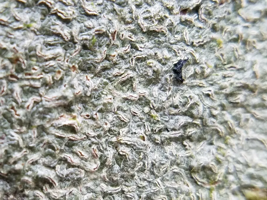
The thallus of Fissurina insidiosa is characterised by its dull grey to dingy olive-grey colour and glossy appearance. Typically, it forms widespread, diffuse patches that can extend up to approximately 10 cm wide. The thallus is usually continuous but often displays cracks and is between 20 and 100 μm thick. It contains crystals of calcium oxalate. The of this species are scattered and typically very numerous. They range in shape from simple to occasionally forked and can be straight, curved, or sinuous, extending up to 2.5 mm in length. Initially, they appear as cracks in the thallus, with the cortex edges curving upwards to form a pseudo-margin. Over time, these develop into a pair of swollen, pale beige-brown lips, which are often cracked and rough, measuring 0.3 to 0.9 mm in width. The of the lirellae remains obscured. The , visible in cross-section, is poorly differentiated from adjacent tissues and measures 10 to 30 μm thick. It has a yellow colour, which reacts K+ (orange-red). The are seldom observed and are approximately 3 μm thick without any warty features. The layer is relatively thin, ranging from 10 to 20 μm in thickness. The hymenium layer is more substantial, measuring between 90 and 120 μm thick. The asci typically contain 6 to 8 spores and measure 85 to 110 by 18 to 25 μm, although intact asci are rarely observed. The paraphyses are slender, about 1 to 1.5 μm wide, with tips that are neither expanded nor adorned with warts or spines. The are broadly ellipsoidal with rounded ends, and they display a transverse 3-septate structure. They measure approximately 13 to 25 by 6 to 9 μm and feature a that can swell when exposed to a solution of potassium hydroxide.

Sebagian uraian ditampilkan dalam bahasa sumber; terjemahannya belum tersedia atau belum lolos pemeriksaan.

Rujukan: [1]

Pengukuran mengikuti objek, spesimen atau kultur, dan metode yang disebutkan sumber. Baca uraian asli untuk konteks lengkap.

02 / Kehidupan

Habitat dan peran ekologis

This species has a broad distribution in the Southern Hemisphere, with recorded presences in mainland Australia, New Zealand, the Pacific region, the Caribbean, and India.

Sebagian uraian ditampilkan dalam bahasa sumber; terjemahannya belum tersedia atau belum lolos pemeriksaan.

Rujukan: [1]

Habitat atau konteks temuan
Informasi belum tersedia pada sumber rilis ini.
Substrat
Informasi belum tersedia pada sumber rilis ini.
Peran atau konteks
Pembentuk liken
Temukan hubungan serupa
03 / Siklus hidup

Reproduksi

The hymenium layer is more substantial, measuring between 90 and 120 μm thick. The asci typically contain 6 to 8 spores and measure 85 to 110 by 18 to 25 μm, although intact asci are rarely observed. The paraphyses are slender, about 1 to 1.5 μm wide, with tips that are neither expanded nor adorned with warts or spines. The are broadly ellipsoidal with rounded ends, and they display a transverse 3-septate structure. They measure approximately 13 to 25 by 6 to 9 μm and feature a that can swell when exposed to a solution of potassium hydroxide. Initially, the of the spores have a lens shape but soon become rounded.

Sebagian uraian ditampilkan dalam bahasa sumber; terjemahannya belum tersedia atau belum lolos pemeriksaan.

Rujukan: [1]

Sinonim menurut acuan taksonomi rilis: Graphis insidiosa
04 / Batas informasi

Catatan identitas dan cakupan

  • Nama diterima mengikuti Catalogue of Life Base2026-09-11; uraian mempertahankan konsep dan nama sumber aslinya.
  • Original geographic, specimen and life-cycle scope retained; source import awaits individual editorial review.
  • Impor dan terjemahan otomatis belum diperiksa editorial per profil. Struktur reproduktif yang disebutkan tidak membuktikan seluruh siklus hidup diketahui.
  • Pengukuran pada uraian mengikuti spesimen, kultur, dan metode sumber; angka tidak diperluas ke semua populasi.

Catatan distribusi dalam narasi mengikuti sumbernya. Dokumentasi spesimen atau foto mengikuti catatan sumber. Bukti wilayah Indonesia dan penilaian konservasi tidak disimpulkan dari gambar.

05 / Dokumentasi

Melihat dengan konteks

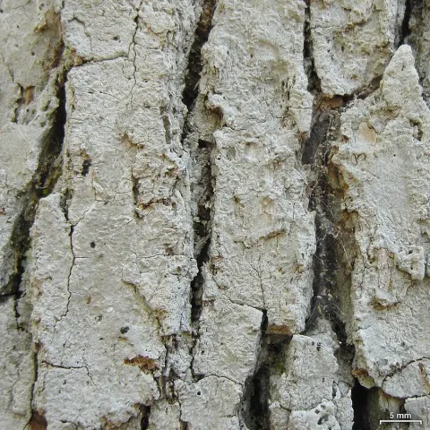
Dokumentasi Fissurina insidiosa menurut asosiasi foto pada record takson iNaturalist. Kredit: Leila Dasher. Identitas mengikuti sumber; bukan penentuan spesimen independen.

Jenis
Fotografi
Kredit
(c) Leila Dasher, some rights reserved (CC BY), uploaded by Leila Dasher
Lisensi
CC BY 4.0
Sumber media
Buka entri sumber

Gambar menunjukkan objek menurut identifikasi sumber, bukan verifikasi independen spesimen oleh Fungi. Tidak ada skala baru yang ditambahkan.

06 / Rujukan

Sumber profil

  1. [1] Fissurina insidiosa — Wikipedia (en)Bagian sumber: Frozen revision 1359162852
    Tanggal penghimpunan sumber: 2026-10-12
    Teks sumber: CC BY-SA4.0 · Wikipedia contributors
  2. [2] Catalogue of Life Base2026-09-11 — Fissurina insidiosaBagian sumber: ColDP NameUsage.tsv ID 6J64G; accepted species; source 2073
    Tanggal penghimpunan sumber: 2026-10-12
  3. [3] Dokumentasi visual — Fissurina insidiosaBagian sumber: Individual media record; identification follows the source, not independent determination
    Tanggal penghimpunan sumber: 2026-10-12
    Media sumber: CC BY 4.0 · (c) Leila Dasher, some rights reserved (CC BY), uploaded by Leila Dasher

Adaptasi teks: CC BY-SA 4.0. Wikipedia contributors — Fissurina insidiosa — Wikipedia (en)

Uraian asli dan riwayat sumber

Riwayat atau halaman sumber

Ringkasan sumber

Fissurina insidiosa is a species of corticolous (bark-dwelling), script lichen in the family Graphidaceae. Found in the Southern Hemisphere, it has been recorded from mainland Australia, New Zealand, the Pacific region, the Caribbean, and India.

Revisi sumber

Morfologi sumber

The thallus of Fissurina insidiosa is characterised by its dull grey to dingy olive-grey colour and glossy appearance. Typically, it forms widespread, diffuse patches that can extend up to approximately 10 cm wide. The thallus is usually continuous but often displays cracks and is between 20 and 100 μm thick. It contains crystals of calcium oxalate. The of this species are scattered and typically very numerous. They range in shape from simple to occasionally forked and can be straight, curved, or sinuous, extending up to 2.5 mm in length. Initially, they appear as cracks in the thallus, with the cortex edges curving upwards to form a pseudo-margin. Over time, these develop into a pair of swollen, pale beige-brown lips, which are often cracked and rough, measuring 0.3 to 0.9 mm in width. The of the lirellae remains obscured. The , visible in cross-section, is poorly differentiated from adjacent tissues and measures 10 to 30 μm thick. It has a yellow colour, which reacts K+ (orange-red). The are seldom observed and are approximately 3 μm thick without any warty features. The layer is relatively thin, ranging from 10 to 20 μm in thickness. The hymenium layer is more substantial, measuring between 90 and 120 μm thick. The asci typically contain 6 to 8 spores and measure 85 to 110 by 18 to 25 μm, although intact asci are rarely observed. The paraphyses are slender, about 1 to 1.5 μm wide, with tips that are neither expanded nor adorned with warts or spines. The are broadly ellipsoidal with rounded ends, and they display a transverse 3-septate structure. They measure approximately 13 to 25 by 6 to 9 μm and feature a that can swell when exposed to a solution of potassium hydroxide.

Revisi sumber

Ekologi sumber

This species has a broad distribution in the Southern Hemisphere, with recorded presences in mainland Australia, New Zealand, the Pacific region, the Caribbean, and India.

Revisi sumber

Struktur reproduksi sumber

The hymenium layer is more substantial, measuring between 90 and 120 μm thick. The asci typically contain 6 to 8 spores and measure 85 to 110 by 18 to 25 μm, although intact asci are rarely observed. The paraphyses are slender, about 1 to 1.5 μm wide, with tips that are neither expanded nor adorned with warts or spines. The are broadly ellipsoidal with rounded ends, and they display a transverse 3-septate structure. They measure approximately 13 to 25 by 6 to 9 μm and feature a that can swell when exposed to a solution of potassium hydroxide. Initially, the of the spores have a lens shape but soon become rounded.

Revisi sumber

Rujukan halaman ini

Fungi. Fissurina insidiosa. Rilis data fungi-2026-10-12-expansion-2. https://fungi-atlas.mgadib.chatgpt.site/jamur/fissurina-insidiosa/.

Status: impor sumber; pemeriksaan editorial per profil belum selesai. Untuk klaim biologis, gunakan rujukan pada bagian terkait dan periksa konteks sumber aslinya.