01 / Morfologi

Ciri dan struktur

Diagnosis: It is characterized by a glossy upper surface, tiny lobes, and a thick algal layer. Description: Thallus squamulose; terricolous; lobes roundish, tiny, 0.7–2 mm wide, (356) 442 ± 55 (523) µm thick; contiguous to densely aggregated, tightly adnate to the substrate; upper surface medium brown, glossy, brown rimmed; lower surface pale to pale yellow; epinecral layer transparent, (33.4) 35.4 ± 1.6 (37.5) µm thick; upper cortex (41) 58 ± 15 (78) µm thick, paraplectenchymatous, cells 8–10 µm in diam.; uppermost layer pale yellow to light brown, 33–36 µm thick; algal layer (104) 159 ± 34 (195) µm thick, algal cells globular, (7.3) 7.8 ± 0.5 (8.7) µm in diam. dispersed over the whole medulla; medulla zone not clear; lower cortex not delimited from the medulla, (42) 63 ± 16 (88) µm thick, composed of densely aggregated globular cells, cells (8.4) 10.6 ± 1.5 (12.6) µm in diam. Rhizohyphae (4.0) 5.2 ± 0.7 (5.9) µm thick, colorless. Pycnidia laminal, immersed, Dermatocarpon -type, immature, subglobular, 250 µm in dim., conidia not seen.

Sebagian uraian ditampilkan dalam bahasa sumber; terjemahannya belum tersedia atau belum lolos pemeriksaan.

Rujukan: [1]

Pengukuran mengikuti objek, spesimen atau kultur, dan metode yang disebutkan sumber. Baca uraian asli untuk konteks lengkap.

02 / Kehidupan

Habitat dan peran ekologis

Ecology and distribution: On the surface of sandy soil in high altitude areas of the Qinghai–Tibet plateau. It has been known only in China up to now.

Sebagian uraian ditampilkan dalam bahasa sumber; terjemahannya belum tersedia atau belum lolos pemeriksaan.

Rujukan: [1]

Habitat atau konteks temuan
Informasi belum tersedia pada sumber rilis ini.
Substrat
Informasi belum tersedia pada sumber rilis ini.
Peran atau konteks
Pembentuk liken
Temukan hubungan serupa
03 / Siklus hidup

Reproduksi

Description: Thallus squamulose; terricolous; lobes roundish, tiny, 0.7–2 mm wide, (356) 442 ± 55 (523) µm thick; contiguous to densely aggregated, tightly adnate to the substrate; upper surface medium brown, glossy, brown rimmed; lower surface pale to pale yellow; epinecral layer transparent, (33.4) 35.4 ± 1.6 (37.5) µm thick; upper cortex (41) 58 ± 15 (78) µm thick, paraplectenchymatous, cells 8–10 µm in diam.; uppermost layer pale yellow to light brown, 33–36 µm thick; algal layer (104) 159 ± 34 (195) µm thick, algal cells globular, (7.3) 7.8 ± 0.5 (8.7) µm in diam. dispersed over the whole medulla; medulla zone not clear; lower cortex not delimited from the medulla, (42) 63 ± 16 (88) µm thick, composed of densely aggregated globular cells, cells (8.4) 10.6 ± 1.5 (12.6) µm in diam.

Sebagian uraian ditampilkan dalam bahasa sumber; terjemahannya belum tersedia atau belum lolos pemeriksaan.

Rujukan: [1]

04 / Batas informasi

Catatan identitas dan cakupan

  • Nama diterima mengikuti Catalogue of Life Base2026-09-11; uraian mempertahankan konsep dan nama sumber aslinya.
  • Preserve original treatment localities, hosts and substrate scope. Materials examined describe documented specimens, not the full global range.
  • Impor dan terjemahan otomatis belum diperiksa editorial per profil. Struktur reproduktif yang disebutkan tidak membuktikan seluruh siklus hidup diketahui.
  • Pengukuran pada uraian mengikuti spesimen, kultur, dan metode sumber; angka tidak diperluas ke semua populasi.

Catatan distribusi dalam narasi mengikuti sumbernya. Dokumentasi spesimen atau foto mengikuti catatan sumber. Bukti wilayah Indonesia dan penilaian konservasi tidak disimpulkan dari gambar.

05 / Dokumentasi

Melihat dengan konteks

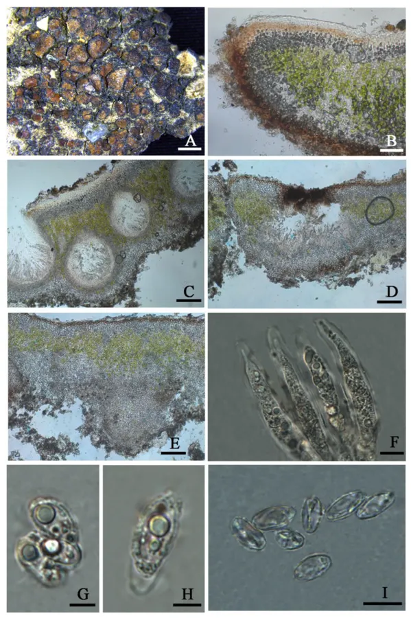
The thallus habit and the anatomic structure of Placidium nitidulum sp. nov (holotype HMAS-L 151947). (A). Squamulose thallus. (B). Transversal section of thallus. (C). Immersed perithecia. (D). Aggregated pyrithecia. (E). Pycnidium. (F). Cylindrical asci with uniseriate ascospores. (G–I). Ascospores. Bars: (A) =2 mm, (B) =50 µm, (C–E) =100 µm, (F) =10 µm, (G, H) =5 µm, and (I) =10 µm.

Jenis
Pelat ilmiah
Kredit
© 2022 by the authors.
Lisensi
CC BY 4.0
Sumber media
Buka entri sumber

Gambar menunjukkan objek menurut identifikasi sumber, bukan verifikasi independen spesimen oleh Fungi. Tidak ada skala baru yang ditambahkan.

06 / Rujukan

Sumber profil

  1. [1] Hidden Species Diversity was Explored in Two Genera of Catapyrenioid Lichens (Verrucariaceae, Ascomycota) from the Deserts of China — Placidium nitidulumBagian sumber: sec3dot2-jof-08-00729_paragraph_25
    Tanggal penghimpunan sumber: 2026-10-12
    Teks sumber: CC BY 4.0 · Zhang T, Zhang X, Yang Q, Wei X.
  2. [2] Catalogue of Life Base2026-09-11 — Placidium nitidulumBagian sumber: ColDP NameUsage.tsv ID B2WNR; accepted species; source 2073
    Tanggal penghimpunan sumber: 2026-10-12
  3. [3] Dokumentasi visual — Placidium nitidulumBagian sumber: Individual media record; identification follows the source, not independent determination
    Tanggal penghimpunan sumber: 2026-10-12
    Media sumber: CC BY 4.0 · © 2022 by the authors.

Adaptasi teks: CC BY 4.0. Zhang T, Zhang X, Yang Q, Wei X. — Hidden Species Diversity was Explored in Two Genera of Catapyrenioid Lichens (Verrucariaceae, Ascomycota) from the Deserts of China — Placidium nitidulum

Uraian asli dan riwayat sumber

Riwayat atau halaman sumber

Ringkasan sumber

Diagnosis: It is characterized by a glossy upper surface, tiny lobes, and a thick algal layer. Description: Thallus squamulose; terricolous; lobes roundish, tiny, 0.7–2 mm wide, (356) 442 ± 55 (523) µm thick; contiguous to densely aggregated, tightly adnate to the substrate; upper surface medium brown, glossy, brown rimmed; lower surface pale to pale yellow; epinecral layer transparent, (33.4) 35.4 ± 1.6 (37.5) µm thick; upper cortex (41) 58 ± 15 (78) µm thick, paraplectenchymatous, cells 8–10 µm in diam.; uppermost layer pale yellow to light brown, 33–36 µm thick; algal layer (104) 159 ± 34 (195) µm thick, algal cells globular, (7.3) 7.8 ± 0.5 (8.7) µm in diam. dispersed over the whole medulla; medulla zone not clear; lower cortex not delimited from the medulla, (42) 63 ± 16 (88) µm thick, composed of densely aggregated globular cells, cells (8.4) 10.6 ± 1.5 (12.6) µm in diam.

Revisi sumber

Morfologi sumber

Diagnosis: It is characterized by a glossy upper surface, tiny lobes, and a thick algal layer. Description: Thallus squamulose; terricolous; lobes roundish, tiny, 0.7–2 mm wide, (356) 442 ± 55 (523) µm thick; contiguous to densely aggregated, tightly adnate to the substrate; upper surface medium brown, glossy, brown rimmed; lower surface pale to pale yellow; epinecral layer transparent, (33.4) 35.4 ± 1.6 (37.5) µm thick; upper cortex (41) 58 ± 15 (78) µm thick, paraplectenchymatous, cells 8–10 µm in diam.; uppermost layer pale yellow to light brown, 33–36 µm thick; algal layer (104) 159 ± 34 (195) µm thick, algal cells globular, (7.3) 7.8 ± 0.5 (8.7) µm in diam. dispersed over the whole medulla; medulla zone not clear; lower cortex not delimited from the medulla, (42) 63 ± 16 (88) µm thick, composed of densely aggregated globular cells, cells (8.4) 10.6 ± 1.5 (12.6) µm in diam. Rhizohyphae (4.0) 5.2 ± 0.7 (5.9) µm thick, colorless. Pycnidia laminal, immersed, Dermatocarpon -type, immature, subglobular, 250 µm in dim., conidia not seen.

Revisi sumber

Ekologi sumber

Ecology and distribution: On the surface of sandy soil in high altitude areas of the Qinghai–Tibet plateau. It has been known only in China up to now.

Revisi sumber

Struktur reproduksi sumber

Description: Thallus squamulose; terricolous; lobes roundish, tiny, 0.7–2 mm wide, (356) 442 ± 55 (523) µm thick; contiguous to densely aggregated, tightly adnate to the substrate; upper surface medium brown, glossy, brown rimmed; lower surface pale to pale yellow; epinecral layer transparent, (33.4) 35.4 ± 1.6 (37.5) µm thick; upper cortex (41) 58 ± 15 (78) µm thick, paraplectenchymatous, cells 8–10 µm in diam.; uppermost layer pale yellow to light brown, 33–36 µm thick; algal layer (104) 159 ± 34 (195) µm thick, algal cells globular, (7.3) 7.8 ± 0.5 (8.7) µm in diam. dispersed over the whole medulla; medulla zone not clear; lower cortex not delimited from the medulla, (42) 63 ± 16 (88) µm thick, composed of densely aggregated globular cells, cells (8.4) 10.6 ± 1.5 (12.6) µm in diam.

Revisi sumber

Rujukan halaman ini

Fungi. Placidium nitidulum. Rilis data fungi-2026-10-12-expansion-2. https://fungi-atlas.mgadib.chatgpt.site/jamur/placidium-nitidulum/.

Status: impor sumber; pemeriksaan editorial per profil belum selesai. Untuk klaim biologis, gunakan rujukan pada bagian terkait dan periksa konteks sumber aslinya.